• 文献检索
  • 文档翻译
  • 深度研究
  • 学术资讯
  • Suppr Zotero 插件Zotero 插件
  • 邀请有礼
  • 套餐&价格
  • 历史记录
应用&插件
Suppr Zotero 插件Zotero 插件浏览器插件Mac 客户端Windows 客户端微信小程序
定价
高级版会员购买积分包购买API积分包
服务
文献检索文档翻译深度研究API 文档MCP 服务
关于我们
关于 Suppr公司介绍联系我们用户协议隐私条款
关注我们

Suppr 超能文献

核心技术专利:CN118964589B侵权必究
粤ICP备2023148730 号-1Suppr @ 2026

文献检索

告别复杂PubMed语法,用中文像聊天一样搜索,搜遍4000万医学文献。AI智能推荐,让科研检索更轻松。

立即免费搜索

文件翻译

保留排版,准确专业,支持PDF/Word/PPT等文件格式,支持 12+语言互译。

免费翻译文档

深度研究

AI帮你快速写综述,25分钟生成高质量综述,智能提取关键信息,辅助科研写作。

立即免费体验

双光子组织成像:以全新视角观察免疫系统。

Two-photon tissue imaging: seeing the immune system in a fresh light.

作者信息

Cahalan Michael D, Parker Ian, Wei Sindy H, Miller Mark J

机构信息

Department of Physiology, University of California, Irvine, California 92697-4561, USA.

出版信息

Nat Rev Immunol. 2002 Nov;2(11):872-80. doi: 10.1038/nri935.

DOI:10.1038/nri935
PMID:12415310
原文链接:https://pmc.ncbi.nlm.nih.gov/articles/PMC2749751/
Abstract

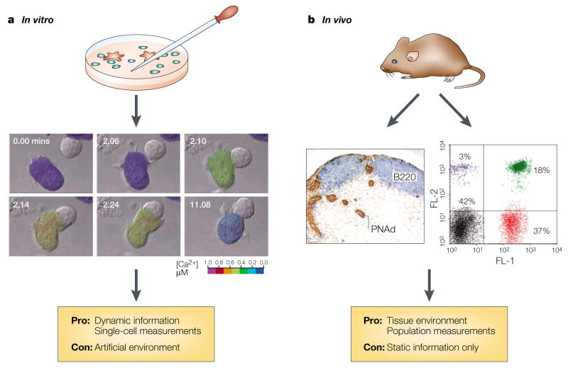
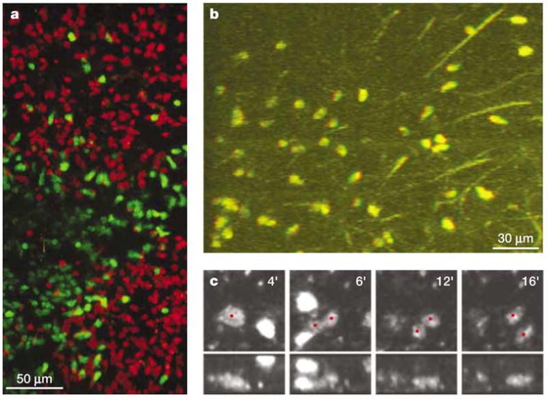
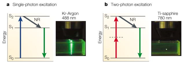
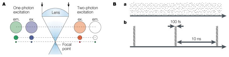
Many lymphocyte functions, such as antigen recognition, take place deep in densely populated lymphoid organs. Because direct in vivo observation was not possible, the dynamics of immune-cell interactions have been inferred or extrapolated from in vitro studies. Two-photon fluorescence excitation uses extremely brief (<1 picosecond) and intense pulses of light to 'see' directly into living tissues, to a greater depth and with less phototoxicity than conventional imaging methods. Two-photon microscopy, in combination with newly developed indicator molecules, promises to extend single-cell approaches to the in vivo setting and to reveal in detail the cellular collaborations that underlie the immune response.

摘要

许多淋巴细胞功能,如抗原识别,发生在人口密集的淋巴器官深处。由于无法进行直接的体内观察,免疫细胞相互作用的动态过程一直是从体外研究中推断或外推出来的。双光子荧光激发使用极短(<1皮秒)且强烈的光脉冲直接“观察”活组织,比传统成像方法能达到更深的深度且光毒性更小。双光子显微镜与新开发的指示分子相结合,有望将单细胞方法扩展到体内环境,并详细揭示免疫反应背后的细胞协作。

https://cdn.ncbi.nlm.nih.gov/pmc/blobs/f2fb/2749751/171c66bb06d0/nihms141989f4.jpg
https://cdn.ncbi.nlm.nih.gov/pmc/blobs/f2fb/2749751/f385c3a53b2a/nihms141989f1.jpg
https://cdn.ncbi.nlm.nih.gov/pmc/blobs/f2fb/2749751/09e781ff3daf/nihms141989f2.jpg
https://cdn.ncbi.nlm.nih.gov/pmc/blobs/f2fb/2749751/13416727cf1f/nihms141989f3.jpg
https://cdn.ncbi.nlm.nih.gov/pmc/blobs/f2fb/2749751/171c66bb06d0/nihms141989f4.jpg

相似文献

1
Two-photon tissue imaging: seeing the immune system in a fresh light.双光子组织成像:以全新视角观察免疫系统。
Nat Rev Immunol. 2002 Nov;2(11):872-80. doi: 10.1038/nri935.
2
Intravital two-photon imaging of T-cell priming and tolerance in the lymph node.淋巴结中T细胞致敏与耐受的活体双光子成像
Methods Mol Biol. 2007;380:355-63. doi: 10.1007/978-1-59745-395-0_22.
3
Two-photon excitation microscopy for the study of living cells and tissues.用于研究活细胞和组织的双光子激发显微镜。
Curr Protoc Cell Biol. 2003 Nov;Chapter 4:Unit 4.11. doi: 10.1002/0471143030.cb0411s20.
4
Live Imaging of Resident T-Cell Migration in Human Lymphoid Tissue Slices Using Confocal Microscopy.使用共聚焦显微镜对人淋巴组织切片中驻留T细胞迁移进行实时成像。
Methods Mol Biol. 2019;1930:75-82. doi: 10.1007/978-1-4939-9036-8_10.
5
Advances in Two-Photon Imaging in Plants.植物双光子成像技术的进展。
Plant Cell Physiol. 2021 Nov 10;62(8):1224-1230. doi: 10.1093/pcp/pcab062.
6
Imaging the choreography of lymphocyte trafficking and the immune response.成像淋巴细胞迁移的编排与免疫反应。
Curr Opin Immunol. 2006 Aug;18(4):476-82. doi: 10.1016/j.coi.2006.05.013. Epub 2006 Jun 12.
7
Molecular photobleaching kinetics of Rhodamine 6G by one- and two-photon induced confocal fluorescence microscopy.罗丹明6G在单光子和双光子诱导共聚焦荧光显微镜下的分子光漂白动力学
Chemphyschem. 2005 May;6(5):791-804. doi: 10.1002/cphc.200400509.
8
An evaluation of two-photon excitation versus confocal and digital deconvolution fluorescence microscopy imaging in Xenopus morphogenesis.非洲爪蟾形态发生过程中双光子激发与共聚焦及数字反卷积荧光显微镜成像的评估。
Microsc Res Tech. 1999 Nov 1;47(3):172-81. doi: 10.1002/(SICI)1097-0029(19991101)47:3<172::AID-JEMT3>3.0.CO;2-A.
9
Simultaneous Two- and Three-Photon Deep Imaging of Autofluorescence in Bacterial Communities.细菌群落中自发荧光的双光子和三光子同步深度成像
Sensors (Basel). 2024 Jan 20;24(2):667. doi: 10.3390/s24020667.
10
Deep tissue two-photon microscopy.深部组织双光子显微镜检查
Nat Methods. 2005 Dec;2(12):932-40. doi: 10.1038/nmeth818.

引用本文的文献

1
Inverse-scattering in biological samples via beam-propagation.通过光束传播对生物样本进行逆散射。
bioRxiv. 2025 Aug 22:2025.08.17.670744. doi: 10.1101/2025.08.17.670744.
2
Mimicking and analyzing the tumor microenvironment.模拟和分析肿瘤微环境。
Cell Rep Methods. 2024 Oct 21;4(10):100866. doi: 10.1016/j.crmeth.2024.100866. Epub 2024 Sep 30.
3
Aggregation-induced emission luminescence for angiography and atherosclerotic diagnosis.用于血管造影和动脉粥样硬化诊断的聚集诱导发光

本文引用的文献

1
Phenotypic analysis of antigen-specific T lymphocytes. Science. 1996. 274: 94-96.抗原特异性T淋巴细胞的表型分析。《科学》。1996年。第274卷:94 - 96页。
J Immunol. 2011 Jul 1;187(1):7-9.
2
Two-photon imaging in intact lymphoid tissue.完整淋巴组织中的双光子成像。
Adv Exp Med Biol. 2002;512:203-8. doi: 10.1007/978-1-4615-0757-4_26.
3
A photoactivatable GFP for selective photolabeling of proteins and cells.一种用于蛋白质和细胞选择性光标记的光激活绿色荧光蛋白。
iScience. 2024 Aug 13;27(9):110719. doi: 10.1016/j.isci.2024.110719. eCollection 2024 Sep 20.
4
Preclinical Models for Cryptococcosis of the CNS and Their Characterization Using In Vivo Imaging Techniques.中枢神经系统隐球菌病的临床前模型及其使用体内成像技术的表征
J Fungi (Basel). 2024 Feb 12;10(2):146. doi: 10.3390/jof10020146.
5
Intravital imaging of the functions of immune cells in the tumor microenvironment during immunotherapy.免疫治疗中肿瘤微环境中免疫细胞功能的活体成像。
Front Immunol. 2023 Dec 6;14:1288273. doi: 10.3389/fimmu.2023.1288273. eCollection 2023.
6
Intravital Microscopy of Lipopolysaccharide-Induced Inflammatory Changes in Different Organ Systems-A Scoping Review.在体显微镜检查不同器官系统中脂多糖诱导的炎症变化:范围综述。
Int J Mol Sci. 2023 Nov 15;24(22):16345. doi: 10.3390/ijms242216345.
7
Two-photon nanoprobes based on bioorganic nanoarchitectonics with a photo-oxidation enhanced emission mechanism.基于生物有机纳架构并具有光氧化增强发射机制的双光子纳米探针。
Nat Commun. 2023 Aug 26;14(1):5227. doi: 10.1038/s41467-023-40897-4.
8
Two-Photon Excitation Spectra of Various Fluorescent Proteins within a Broad Excitation Range.在宽激发范围内的各种荧光蛋白的双光子激发光谱。
Int J Mol Sci. 2022 Nov 2;23(21):13407. doi: 10.3390/ijms232113407.
9
Phosphorescent Ir(III) Complexes for Biolabeling and Biosensing.用于生物标记和生物传感的磷光铱(III)配合物。
Top Curr Chem (Cham). 2022 Aug 10;380(5):35. doi: 10.1007/s41061-022-00389-3.
10
Visualization and characterization of metallo-aggregates using multi-photon microscopy.使用多光子显微镜对金属聚集体进行可视化和表征。
RSC Adv. 2020 Dec 24;11(2):657-661. doi: 10.1039/d0ra07263j.
Science. 2002 Sep 13;297(5588):1873-7. doi: 10.1126/science.1074952.
4
Chemokine requirements for B cell entry to lymph nodes and Peyer's patches.B细胞进入淋巴结和派尔集合淋巴结所需的趋化因子
J Exp Med. 2002 Jul 1;196(1):65-75. doi: 10.1084/jem.20020201.
5
Dynamics of thymocyte-stromal cell interactions visualized by two-photon microscopy.通过双光子显微镜观察到的胸腺细胞与基质细胞相互作用的动态过程。
Science. 2002 Jun 7;296(5574):1876-80. doi: 10.1126/science.1070945.
6
Dynamic imaging of T cell-dendritic cell interactions in lymph nodes.淋巴结中T细胞与树突状细胞相互作用的动态成像
Science. 2002 Jun 7;296(5574):1873-6. doi: 10.1126/science.1071065.
7
Immunology. T cell activation in six dimensions.免疫学。六个维度的T细胞活化
Science. 2002 Jun 7;296(5574):1815-7. doi: 10.1126/science.296.5574.1815.
8
Two-photon imaging of lymphocyte motility and antigen response in intact lymph node.完整淋巴结中淋巴细胞运动性和抗原反应的双光子成像
Science. 2002 Jun 7;296(5574):1869-73. doi: 10.1126/science.1070051. Epub 2002 May 16.
9
Inhibition of T cell receptor-coreceptor interactions by antagonist ligands visualized by live FRET imaging of the T-hybridoma immunological synapse.通过T杂交瘤免疫突触的实时荧光共振能量转移成像观察拮抗剂配体对T细胞受体-共受体相互作用的抑制作用。
Immunity. 2002 Apr;16(4):521-34. doi: 10.1016/s1074-7613(02)00301-1.
10
Control of leukocyte rolling velocity in TNF-alpha-induced inflammation by LFA-1 and Mac-1.通过淋巴细胞功能相关抗原-1(LFA-1)和巨噬细胞抗原-1(Mac-1)对肿瘤坏死因子-α(TNF-α)诱导炎症中白细胞滚动速度的调控
Blood. 2002 Jan 1;99(1):336-41. doi: 10.1182/blood.v99.1.336.