Chipev Constantin C, Simon Marcia
Living Skin Bank, University Hospital, Dept Oral Biology and Pathology, HSC, SUNY at Stony Brook, NY 11794-9702, USA.
BMC Dermatol. 2002 Nov 21;2:13. doi: 10.1186/1471-5945-2-13.
Wounds in the nonglabrous skin of keloid-prone individuals tend to cause large disordered accumulations of collagen which extend beyond the original margins of the wound. In addition to abnormalities in keloid fibroblasts, comparison of dermal fibroblasts derived from nonwounded glabrous or nonglabrous skin revealed differences that may account for the observed location of keloids.
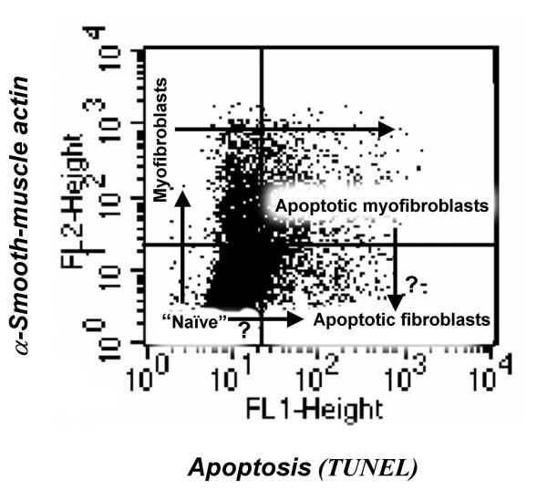
Fibroblast apoptosis and the cellular content of alpha-smooth-muscle actin, TGFbeta1 receptorII and ED-A fibronectin were estimated by FACS analysis. The effects of TGFbeta1 and serum were examined.
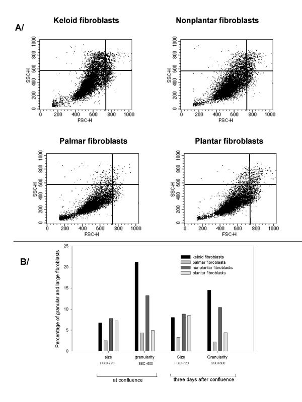
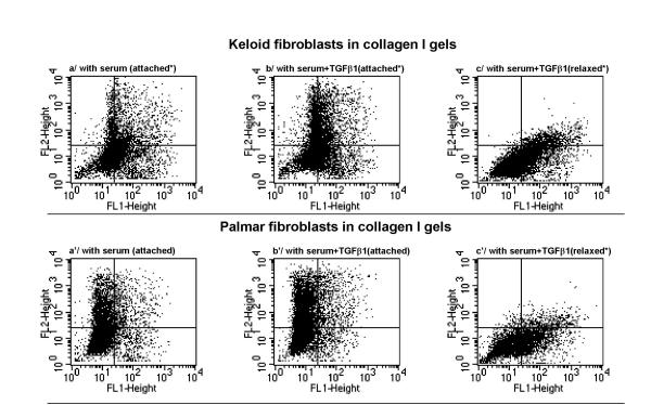
In monolayer cultures non-glabrous fibroblasts were slower growing, had higher granularity and accumulated more alpha-smooth-muscle actin than fibroblasts from glabrous tissues. Keloid fibroblasts had the highest level of alpha-smooth-muscle actin in parallel with their expression level of ED-A fibronectin. TGFbeta1 positively regulated alpha-smooth-muscle actin expression in all fibroblast cultures, although its effects on apoptosis in fibroblasts from glabrous and non-glabrous tissues were found to differ. The presence of collagen I in the ECM resulted in reduction of alpha-smooth-muscle actin. A considerable percentage of the apoptotic fibroblasts in attached gels were alpha-smooth-muscle actin positive. The extent of apoptosis correlated positively with increased cell and matrix relaxation. TGFbeta1 was unable to overcome this apoptotic effect of matrix relaxation.
The presence of myofibroblasts and the apoptosis level can be regulated by both TGFbeta1 and by the extracellular matrix. However, reduction of tension in the matrix is the critical determinant. This predicts that the tension in the wound bed determines the type of scar at different body sites.
瘢痕疙瘩易患个体的非无毛皮肤伤口往往会导致大量无序的胶原蛋白堆积,超出伤口的原始边缘。除了瘢痕疙瘩成纤维细胞的异常外,对来自未受伤无毛或非无毛皮肤的真皮成纤维细胞进行比较,发现了一些差异,这些差异可能解释了观察到的瘢痕疙瘩的位置。
通过流式细胞术分析估计成纤维细胞凋亡以及α-平滑肌肌动蛋白、转化生长因子β1受体II和ED-A纤连蛋白的细胞含量。研究了转化生长因子β1和血清的作用。
在单层培养中,非无毛成纤维细胞生长较慢,颗粒度较高,比来自无毛组织的成纤维细胞积累更多的α-平滑肌肌动蛋白。瘢痕疙瘩成纤维细胞的α-平滑肌肌动蛋白水平最高,与其ED-A纤连蛋白的表达水平平行。转化生长因子β1在所有成纤维细胞培养物中均正向调节α-平滑肌肌动蛋白的表达,尽管发现其对无毛和非无毛组织来源的成纤维细胞凋亡的影响有所不同。细胞外基质中I型胶原蛋白的存在导致α-平滑肌肌动蛋白减少。附着凝胶中相当比例的凋亡成纤维细胞为α-平滑肌肌动蛋白阳性。凋亡程度与细胞和基质松弛增加呈正相关。转化生长因子β1无法克服基质松弛的这种凋亡作用。
肌成纤维细胞的存在和凋亡水平可由转化生长因子β1和细胞外基质共同调节。然而,基质张力的降低是关键决定因素。这预示着伤口床的张力决定了不同身体部位瘢痕的类型。