• 文献检索
  • 文档翻译
  • 深度研究
  • 学术资讯
  • Suppr Zotero 插件Zotero 插件
  • 邀请有礼
  • 套餐&价格
  • 历史记录
应用&插件
Suppr Zotero 插件Zotero 插件浏览器插件Mac 客户端Windows 客户端微信小程序
定价
高级版会员购买积分包购买API积分包
服务
文献检索文档翻译深度研究API 文档MCP 服务
关于我们
关于 Suppr公司介绍联系我们用户协议隐私条款
关注我们

Suppr 超能文献

核心技术专利:CN118964589B侵权必究
粤ICP备2023148730 号-1Suppr @ 2026

文献检索

告别复杂PubMed语法,用中文像聊天一样搜索,搜遍4000万医学文献。AI智能推荐,让科研检索更轻松。

立即免费搜索

文件翻译

保留排版,准确专业,支持PDF/Word/PPT等文件格式,支持 12+语言互译。

免费翻译文档

深度研究

AI帮你快速写综述,25分钟生成高质量综述,智能提取关键信息,辅助科研写作。

立即免费体验

相似文献

1
Multiple signaling pathways converge on the Cbfa1/Runx2 transcription factor to regulate osteoblast differentiation.多条信号通路汇聚于Cbfa1/Runx2转录因子以调控成骨细胞分化。
Connect Tissue Res. 2003;44 Suppl 1(Suppl 1):109-16.
2
Regulation of the osteoblast-specific transcription factor, Runx2: responsiveness to multiple signal transduction pathways.成骨细胞特异性转录因子Runx2的调控:对多种信号转导途径的反应性
J Cell Biochem. 2003 Feb 15;88(3):446-54. doi: 10.1002/jcb.10369.
3
Parathyroid hormone-related peptide (PTHrP) inhibits Runx2 expression through the PKA signaling pathway.甲状旁腺激素相关肽(PTHrP)通过蛋白激酶A(PKA)信号通路抑制Runx2表达。
Exp Cell Res. 2004 Sep 10;299(1):128-36. doi: 10.1016/j.yexcr.2004.05.025.
4
Identification of potential modifiers of Runx2/Cbfa1 activity in C2C12 cells in response to bone morphogenetic protein-7.鉴定C2C12细胞中响应骨形态发生蛋白-7时Runx2/Cbfa1活性的潜在调节因子。
Cells Tissues Organs. 2004;176(1-3):28-40. doi: 10.1159/000075025.
5
Both the Smad and p38 MAPK pathways play a crucial role in Runx2 expression following induction by transforming growth factor-beta and bone morphogenetic protein.在转化生长因子-β和骨形态发生蛋白诱导后,Smad和p38丝裂原活化蛋白激酶(MAPK)信号通路在Runx2表达中均发挥关键作用。
Oncogene. 2002 Oct 17;21(47):7156-63. doi: 10.1038/sj.onc.1205937.
6
Differential regulation of the two principal Runx2/Cbfa1 n-terminal isoforms in response to bone morphogenetic protein-2 during development of the osteoblast phenotype.在成骨细胞表型发育过程中,两种主要的Runx2/Cbfa1 N端亚型对骨形态发生蛋白-2的差异调节。
Endocrinology. 2001 Sep;142(9):4026-39. doi: 10.1210/endo.142.9.8367.
7
Growth hormone attenuates the transcriptional activity of Runx2 by facilitating its physical association with Stat3beta.生长激素通过促进Runx2与Stat3β的物理结合来减弱其转录活性。
J Bone Miner Res. 2004 Nov;19(11):1892-904. doi: 10.1359/JBMR.040701. Epub 2004 Jul 7.
8
Establishment and characterization of a stable cell line to evaluate cellular Runx2 activity.
J Cell Biochem. 2004 Apr 15;91(6):1239-47. doi: 10.1002/jcb.20038.
9
Smad function and intranuclear targeting share a Runx2 motif required for osteogenic lineage induction and BMP2 responsive transcription.Smad功能与核内靶向作用共享一个成骨谱系诱导和BMP2反应性转录所需的Runx2基序。
J Cell Physiol. 2005 Jul;204(1):63-72. doi: 10.1002/jcp.20258.
10
Reconstitution of Runx2/Cbfa1-null cells identifies a requirement for BMP2 signaling through a Runx2 functional domain during osteoblast differentiation.Runx2/Cbfa1基因敲除细胞的重组表明,在成骨细胞分化过程中,通过Runx2功能域的骨形态发生蛋白2(BMP2)信号传导是必需的。
J Cell Biochem. 2007 Feb 1;100(2):434-49. doi: 10.1002/jcb.21039.

引用本文的文献

1
Molecular Determinants of Bone Plasticity Regeneration After Trauma: Forensic Consequences.创伤后骨可塑性再生的分子决定因素:法医学后果
Int J Mol Sci. 2025 Jul 25;26(15):7184. doi: 10.3390/ijms26157184.
2
Caveolin-1 Regulates Parathyroid Hormone (PTH)-Related Protein (PTHrP) Actions on PTH Receptor Type 1 in Bone Cells.小窝蛋白-1调节甲状旁腺激素(PTH)相关蛋白(PTHrP)对骨细胞中1型PTH受体的作用。
J Cell Physiol. 2025 Jul;240(7):e70067. doi: 10.1002/jcp.70067.
3
The Influence of Different Preservation Protocols on the Teeth's Osteoinductive Characteristics: An In Vitro Study.不同保存方案对牙齿骨诱导特性的影响:一项体外研究
Int J Mol Sci. 2025 Apr 24;26(9):4044. doi: 10.3390/ijms26094044.
4
Epigallocatechin gallate protects MC3T3-E1 cells from cadmium-induced apoptosis and dysfunction via modulating PI3K/AKT/mTOR and Nrf2/HO-1 pathways.没食子酸表没食子儿茶素酯通过调控 PI3K/AKT/mTOR 和 Nrf2/HO-1 通路保护 MC3T3-E1 细胞免受镉诱导的凋亡和功能障碍。
PeerJ. 2024 May 28;12:e17488. doi: 10.7717/peerj.17488. eCollection 2024.
5
N6-Methyladenosine in Cell-Fate Determination of BMSCs: From Mechanism to Applications.N6-甲基腺苷在骨髓间充质干细胞命运决定中的作用:从机制到应用
Research (Wash D C). 2024 Apr 25;7:0340. doi: 10.34133/research.0340. eCollection 2024.
6
Biological Evaluation of the Osteoinductive Potential of Dry Teeth after Chemical Demineralization Treatment Using the Tooth Transformer Device.利用牙变形装置对化学脱矿处理后的干牙进行骨诱导潜力的生物学评价。
Biomolecules. 2023 Nov 30;13(12):1727. doi: 10.3390/biom13121727.
7
Inhibition of focal adhesion kinase 2 results in a macrophage polarization shift to M2 which attenuates local and systemic inflammation and reduces heterotopic ossification after polysystem extremity trauma.抑制黏着斑激酶 2 可导致巨噬细胞向 M2 极化转变,从而减轻局部和全身炎症反应,并减少多系统肢体创伤后的异位骨化。
Front Immunol. 2023 Dec 5;14:1280884. doi: 10.3389/fimmu.2023.1280884. eCollection 2023.
8
Sword Bean () Pods Induce Differentiation in MC3T3-E1 Osteoblast Cells by Activating the BMP2/SMAD/RUNX2 Pathway.刀豆()荚果通过激活 BMP2/SMAD/RUNX2 通路诱导 MC3T3-E1 成骨细胞分化。
Nutrients. 2023 Oct 16;15(20):4372. doi: 10.3390/nu15204372.
9
The Interplay of Raloxifene and Sonochemical Bio-Oss in Early Maxillary Sinus Bone Regeneration: A Histological and Immunohistochemical Analysis in Rabbits.雷洛昔芬与超声生物骨在早期上颌窦骨再生中的相互作用:兔的组织学和免疫组织化学分析。
Medicina (Kaunas). 2023 Aug 23;59(9):1521. doi: 10.3390/medicina59091521.
10
Osteoarthritis: pathogenic signaling pathways and therapeutic targets.骨关节炎:发病信号通路和治疗靶点。
Signal Transduct Target Ther. 2023 Feb 3;8(1):56. doi: 10.1038/s41392-023-01330-w.

本文引用的文献

1
Bone morphogenetic proteins, extracellular matrix, and mitogen-activated protein kinase signaling pathways are required for osteoblast-specific gene expression and differentiation in MC3T3-E1 cells.骨形态发生蛋白、细胞外基质和丝裂原活化蛋白激酶信号通路是MC3T3-E1细胞中骨细胞特异性基因表达和分化所必需的。
J Bone Miner Res. 2002 Jan;17(1):101-10. doi: 10.1359/jbmr.2002.17.1.101.
2
Physical interaction of the activator protein-1 factors c-Fos and c-Jun with Cbfa1 for collagenase-3 promoter activation.激活蛋白-1因子c-Fos和c-Jun与Cbfa1在胶原酶-3启动子激活过程中的物理相互作用。
J Biol Chem. 2002 Jan 4;277(1):816-22. doi: 10.1074/jbc.M107082200. Epub 2001 Oct 18.
3
AP-1 and Cbfa/runt physically interact and regulate parathyroid hormone-dependent MMP13 expression in osteoblasts through a new osteoblast-specific element 2/AP-1 composite element.AP-1与Cbfa/runt发生物理相互作用,并通过一种新的成骨细胞特异性元件2/AP-1复合元件调控成骨细胞中甲状旁腺激素依赖性MMP13的表达。
J Biol Chem. 2001 Jun 8;276(23):20029-38. doi: 10.1074/jbc.M010601200. Epub 2001 Mar 26.
4
Cbfa1 contributes to the osteoblast-specific expression of type I collagen genes.Cbfa1 有助于 I 型胶原基因的成骨细胞特异性表达。
J Biol Chem. 2001 Mar 9;276(10):7101-7. doi: 10.1074/jbc.M006215200. Epub 2000 Dec 5.
5
Identification of a novel response element in the rat bone sialoprotein (BSP) gene promoter that mediates constitutive and fibroblast growth factor 2-induced expression of BSP.
J Biol Chem. 2001 Feb 23;276(8):5459-66. doi: 10.1074/jbc.M008971200. Epub 2000 Nov 21.
6
Runx2 is a common target of transforming growth factor beta1 and bone morphogenetic protein 2, and cooperation between Runx2 and Smad5 induces osteoblast-specific gene expression in the pluripotent mesenchymal precursor cell line C2C12.Runx2是转化生长因子β1和骨形态发生蛋白2的共同靶点,Runx2与Smad5之间的协同作用可在多能间充质前体细胞系C2C12中诱导成骨细胞特异性基因表达。
Mol Cell Biol. 2000 Dec;20(23):8783-92. doi: 10.1128/MCB.20.23.8783-8792.2000.
7
The osteoblast: a sophisticated fibroblast under central surveillance.成骨细胞:受中枢监测的复杂成纤维细胞。
Science. 2000 Sep 1;289(5484):1501-4. doi: 10.1126/science.289.5484.1501.
8
A Pro250Arg substitution in mouse Fgfr1 causes increased expression of Cbfa1 and premature fusion of calvarial sutures.小鼠Fgfr1中的Pro250Arg替代导致Cbfa1表达增加和颅缝过早融合。
Hum Mol Genet. 2000 Aug 12;9(13):2001-8. doi: 10.1093/hmg/9.13.2001.
9
FGF receptor mutations: dimerization syndromes, cell growth suppression, and animal models.成纤维细胞生长因子受体突变:二聚化综合征、细胞生长抑制及动物模型
IUBMB Life. 2000 Mar;49(3):197-205. doi: 10.1080/713803609.
10
The developmental control of osteoblast-specific gene expression: role of specific transcription factors and the extracellular matrix environment.成骨细胞特异性基因表达的发育控制:特定转录因子和细胞外基质环境的作用。
Crit Rev Oral Biol Med. 1999;10(1):40-57. doi: 10.1177/10454411990100010201.

多条信号通路汇聚于Cbfa1/Runx2转录因子以调控成骨细胞分化。

Multiple signaling pathways converge on the Cbfa1/Runx2 transcription factor to regulate osteoblast differentiation.

作者信息

Franceschi Renny T, Xiao Guozhi, Jiang Di, Gopalakrishnan Rajaram, Yang Shuying, Reith Elizabeth

机构信息

Department of Periodontics/Prevention/Geriatrics, School of Medicine, University of Michigan, Ann Arbor, Michigan 48109-1078, USA.

出版信息

Connect Tissue Res. 2003;44 Suppl 1(Suppl 1):109-16.

PMID:12952183
原文链接:https://pmc.ncbi.nlm.nih.gov/articles/PMC3564252/
Abstract

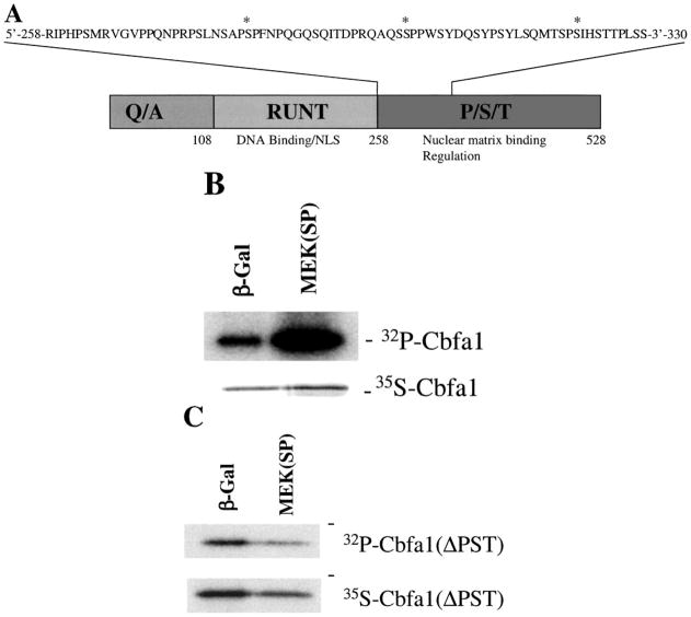
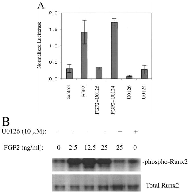
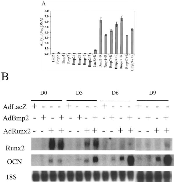
The Cbfa1/Runx2 transcription factor is essential for osteoblast differentiation. However, levels of Runx2 are often not well correlated with its transcriptional activity suggesting that this factor must be activated either by covalent modification or through interactions with other nuclear components. Runx2 is phosphorylated and activated by the mitogen-activated protein kinase (MAPK) pathway. This pathway is stimulated in at least two ways: by binding of type I collagen to alpha2beta1 integrins on the osteoblast surface and by treatment of cells with the osteogenic growth factor, FGF2. Protein kinase A (PKA) also may phosphorylate/activate Runx2 under certain conditions. Runx2 activity also is enhanced by factors known to stimulate specific signal transduction pathways such as PTH/PTHrP (signals through PKA and PKC pathways) and BMPs (Signal through Smad proteins). Interactions with Runx2 are complex involving both binding of distinct components such as AP-1 factors and Smads to separate sites on DNA, direct interactions between Runx2 and AP-1/Smad factors and MAPK or PKA-dependent Runx2 phosphorylation. These findings suggest that Runx2 plays a central role in coordinating multiple signals involved in osteoblast differentiation.

摘要

Cbfa1/Runx2转录因子对成骨细胞分化至关重要。然而,Runx2的水平与其转录活性往往没有很好的相关性,这表明该因子必须通过共价修饰或与其他核成分相互作用才能被激活。Runx2可被丝裂原活化蛋白激酶(MAPK)途径磷酸化并激活。该途径至少通过两种方式被刺激:通过I型胶原与成骨细胞表面的α2β1整合素结合,以及用成骨生长因子FGF2处理细胞。蛋白激酶A(PKA)在某些条件下也可能使Runx2磷酸化/激活。已知刺激特定信号转导途径的因子,如甲状旁腺激素/甲状旁腺激素相关蛋白(通过PKA和PKC途径发出信号)和骨形态发生蛋白(通过Smad蛋白发出信号),也可增强Runx2的活性。与Runx2的相互作用很复杂,涉及不同成分(如AP-1因子和Smads)与DNA上不同位点的结合、Runx2与AP-1/Smad因子之间的直接相互作用以及MAPK或PKA依赖的Runx2磷酸化。这些发现表明,Runx2在协调成骨细胞分化中涉及的多种信号方面发挥着核心作用。

https://cdn.ncbi.nlm.nih.gov/pmc/blobs/1c83/3564252/5bc078124b11/nihms435865f4.jpg
https://cdn.ncbi.nlm.nih.gov/pmc/blobs/1c83/3564252/6f5a5c789240/nihms435865f1.jpg
https://cdn.ncbi.nlm.nih.gov/pmc/blobs/1c83/3564252/a95892c15a8c/nihms435865f2.jpg
https://cdn.ncbi.nlm.nih.gov/pmc/blobs/1c83/3564252/1c0f091167b8/nihms435865f3.jpg
https://cdn.ncbi.nlm.nih.gov/pmc/blobs/1c83/3564252/5bc078124b11/nihms435865f4.jpg