Casanova M, Lopez-Ribot J L, Martinez J P, Sentandreu R
Sección Departamental de Microbiología, Facultad de Farmacia, Universitat de València, Spain.
Infect Immun. 1992 Nov;60(11):4898-906. doi: 10.1128/iai.60.11.4898-4906.1992.
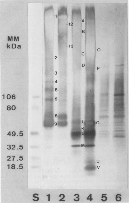
Candida albicans ATCC 26555 blastoconidia and blastoconidia bearing germ tubes were metabolically labelled by incubating the cells with 14C-labelled protein hydrolysate and were subsequently tagged with biotin. Double-labelled (radioactive and biotinylated) cell wall proteins and glycoproteins were extracted from intact cells of both growth forms by treatment with 2-mercaptoethanol (beta ME) and with beta-glucanases (Zymolyase) after treatment with beta ME. The beta ME- and Zymolyase-extracts were separated by sodium dodecyl sulfate-polyacrylamide gel electrophoresis and western blotted (immunoblotted) to nitrocellulose paper. Polyacrylamide gels were stained with Coomassie blue and processed for fluorography. Western blot analysis was performed either with peroxidase conjugated-concanavalin A (ConA) or Extravidin. Blotted proteins were also reacted with polyclonal antibodies and monoclonal antibodies against mannoprotein components from mycelial cell walls of the ATCC 26555 strain. Labelling with biotin allowed identification of a complex array of cell wall protein and glycoprotein components within a very wide molecular mass range (from 650 to 13 kDa). These appeared to be genuine cell wall components. Biotinylated high-molecular-mass glycoproteins that were not stained with Coomassie blue or that appeared as poorly resolved polydisperse bands by indirect ConA-peroxidase staining of Western blots were detected as sharply defined bands following reaction with the Extravidin-peroxidase conjugate. Biotinylated molecules retained unaltered reactivities against ConA, polyclonal antibodies, and monoclonal antibodies.
将白色念珠菌ATCC 26555的芽生孢子和带有芽管的芽生孢子与14C标记的蛋白水解物一起孵育进行代谢标记,随后用生物素进行标记。通过用2-巯基乙醇(βME)处理,然后在用βME处理后用β-葡聚糖酶(溶壁酶)处理,从两种生长形式的完整细胞中提取双标记(放射性和生物素化)的细胞壁蛋白和糖蛋白。βME提取物和溶壁酶提取物通过十二烷基硫酸钠-聚丙烯酰胺凝胶电泳进行分离,并转移至硝酸纤维素纸上进行蛋白质免疫印迹(免疫印迹)。聚丙烯酰胺凝胶用考马斯亮蓝染色并进行荧光自显影处理。蛋白质免疫印迹分析使用过氧化物酶偶联的伴刀豆球蛋白A(ConA)或抗生物素蛋白进行。印迹蛋白还与针对ATCC 26555菌株菌丝细胞壁甘露糖蛋白成分的多克隆抗体和单克隆抗体反应。用生物素标记可以鉴定出分子量范围非常宽(从650至13 kDa)的一系列复杂的细胞壁蛋白和糖蛋白成分。这些似乎是真正的细胞壁成分。在与抗生物素蛋白-过氧化物酶偶联物反应后,未被考马斯亮蓝染色或在蛋白质免疫印迹的间接ConA-过氧化物酶染色中表现为分辨率差的多分散条带的生物素化高分子量糖蛋白被检测为清晰定义的条带。生物素化分子对ConA、多克隆抗体和单克隆抗体的反应性保持不变。