Cromwell O, Hamid Q, Corrigan C J, Barkans J, Meng Q, Collins P D, Kay A B
Department of Allergy and Clinical Immunology, National Heart and Lung Institute, London.
Immunology. 1992 Nov;77(3):330-7.
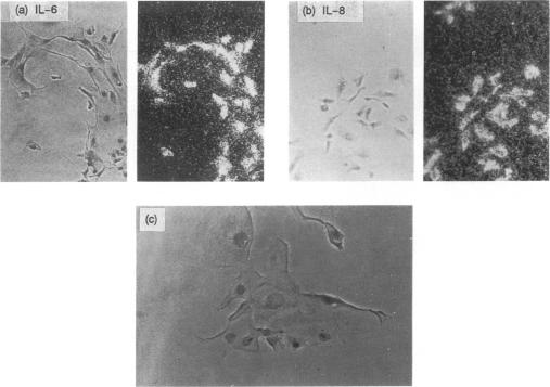
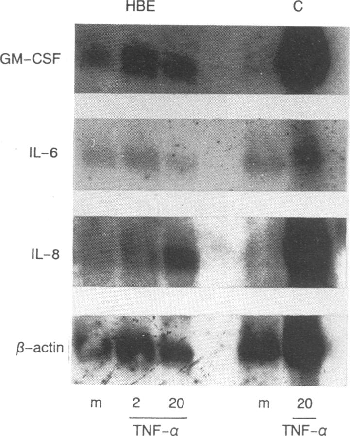
We have tested the hypothesis that the bronchial epithelium has the capacity to generate and release cytokines that could contribute to inflammatory events associated with inflammatory lung diseases. Messenger RNA (mRNA) for interleukin-6 (IL-6), IL-8 and granulocyte-macrophage colony-stimulating factor (GM-CSF) was identified in human bronchial epithelial cell primary cultures, characterized on the basis of staining for cytokeratin, using both in situ hybridization and Northern blotting. Using in situ hybridization we have shown that the majority of the cells expressed mRNA for IL-6 and IL-8, whereas fewer than 20% of cells expressed message for GM-CSF. The numbers of cells expressing message were increased by culture with tumour necrosis factor-alpha (TNF-alpha) (20 ng/ml, 24 hr). These observations were substantiated by Northern blotting, which showed that both TNF-alpha and IL-1 beta were able to induce a dose-dependent increase in IL-8-specific mRNA. Immunoreactive IL-6 and GM-CSF were detected and quantified in the culture supernatants by ELISA, and IL-8 by radioimmunoassay. The levels of immunoreactivity were increased by incubation of epithelial cells with either IL-1 beta or TNF-alpha for 24 hr. A transformed tracheal epithelial cell line (9HTEo-) expressed mRNA for IL-6, IL-8 and GM-CSF but, whereas levels of immunoreactive IL-6 in culture supernatants were comparable with those in primary cell cultures, levels of IL-8 were low and GM-CSF trivial. These observations indicate that the bronchial epithelium has the potential to be a major source of IL-8 and a number of other cytokines, and that production can be amplified substantially by IL-1 beta and TNF-alpha. The bronchial epithelium is ideally situated to modulate inflammatory and immunological events in and around the airways, and these observations suggest that it could contribute to promote and sustain inflammatory and immunological processes in inflammatory lung diseases such asthma.
支气管上皮具有生成和释放细胞因子的能力,这些细胞因子可能促成与炎症性肺病相关的炎症反应。在人支气管上皮细胞原代培养物中鉴定出了白细胞介素-6(IL-6)、IL-8和粒细胞-巨噬细胞集落刺激因子(GM-CSF)的信使核糖核酸(mRNA),通过细胞角蛋白染色、原位杂交和Northern印迹法对其进行了表征。通过原位杂交我们发现,大多数细胞表达IL-6和IL-8的mRNA,而表达GM-CSF信使的细胞不到20%。用肿瘤坏死因子-α(TNF-α)(20纳克/毫升,24小时)培养可增加表达信使的细胞数量。Northern印迹法证实了这些观察结果,该方法显示TNF-α和IL-1β均能诱导IL-8特异性mRNA呈剂量依赖性增加。通过酶联免疫吸附测定法(ELISA)检测并定量培养上清液中的免疫反应性IL-6和GM-CSF,通过放射免疫测定法检测IL-8。用IL-1β或TNF-α孵育上皮细胞24小时可增加免疫反应性水平。一种转化的气管上皮细胞系(9HTEo-)表达IL-6、IL-8和GM-CSF的mRNA,但是,尽管培养上清液中免疫反应性IL-6的水平与原代细胞培养物中的相当,但IL-8的水平较低,GM-CSF的水平微不足道。这些观察结果表明,支气管上皮有可能成为IL-8和许多其他细胞因子的主要来源,并且IL-1β和TNF-α可大幅放大其产生。支气管上皮处于调节气道内及周围炎症和免疫反应的理想位置,这些观察结果表明,它可能有助于促进和维持诸如哮喘等炎症性肺病中的炎症和免疫过程。