Suárez-Fariñas Mayte, Haider Asifa, Wittkowski Knut M
Center for Studies in Physics and Biology, The Rockefeller University, 1230 York Ave, Box 212, New York, NY 10021, USA.
BMC Bioinformatics. 2005 Mar 22;6:65. doi: 10.1186/1471-2105-6-65.
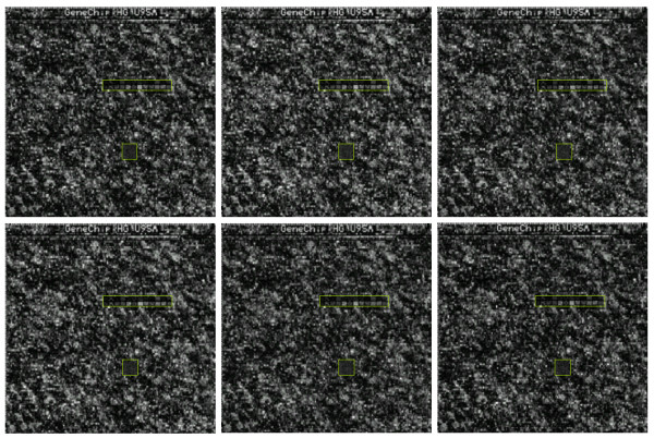
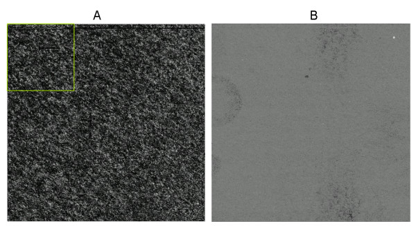
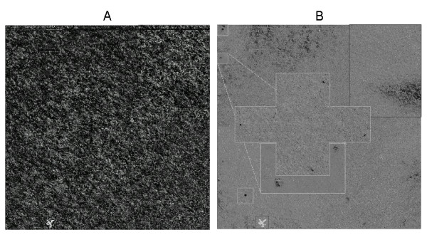
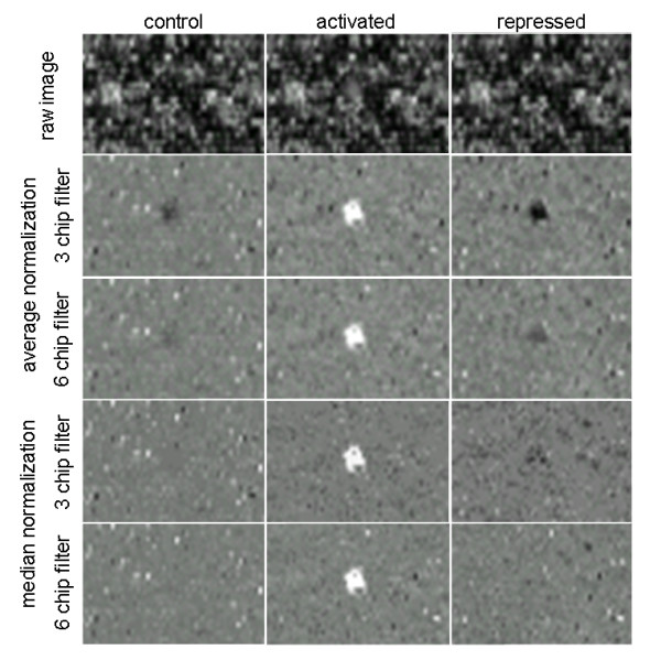
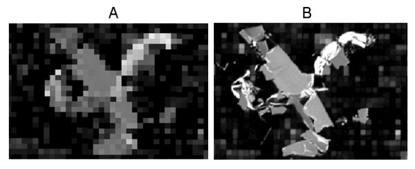
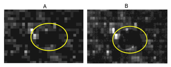
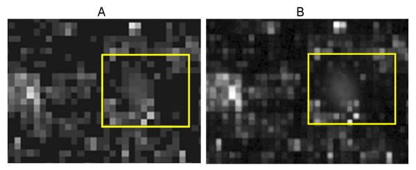
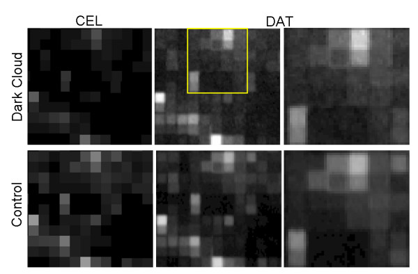
Microscopists are familiar with many blemishes that fluorescence images can have due to dust and debris, glass flaws, uneven distribution of fluids or surface coatings, etc. Microarray scans show similar artefacts, which affect the analysis, particularly when one tries to detect subtle changes. However, most blemishes are hard to find by the unaided eye, particularly in high-density oligonucleotide arrays (HDONAs).
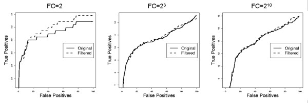
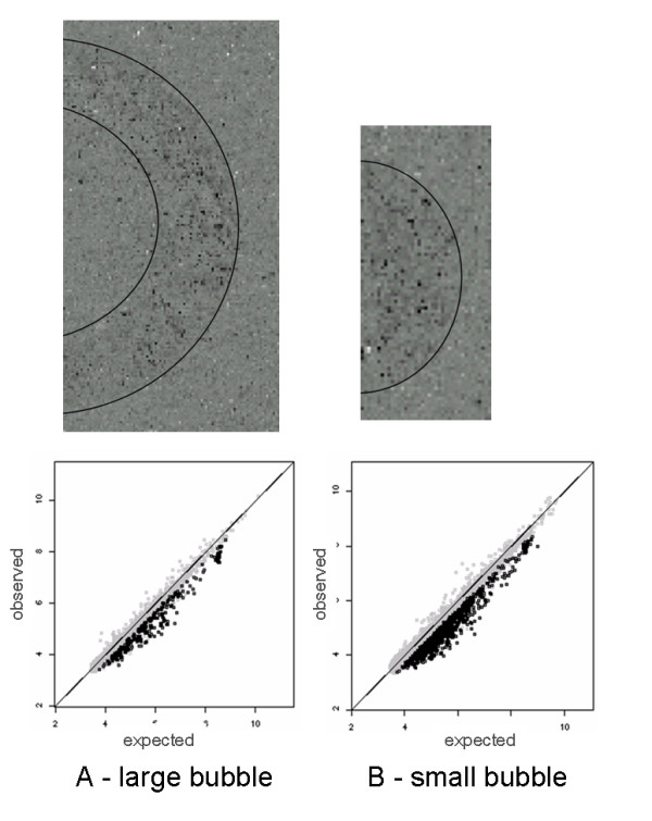
We present a method that harnesses the statistical power provided by having several HDONAs available, which are obtained under similar conditions except for the experimental factor. This method "harshlights" blemishes and renders them evident. We find empirically that about 25% of our chips are blemished, and we analyze the impact of masking them on screening for differentially expressed genes.
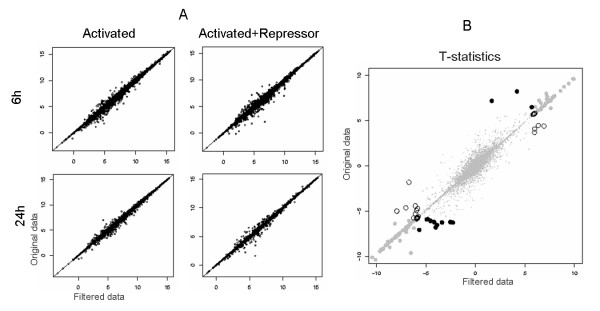
Experiments attempting to assess subtle expression changes should be carefully screened for blemishes on the chips. The proposed method provides investigators with a novel robust approach to improve the sensitivity of microarray analyses. By utilizing topological information to identify and mask blemishes prior to model based analyses, the method prevents artefacts from confounding the process of background correction, normalization, and summarization.
显微镜学家熟知荧光图像可能因灰尘和碎屑、玻璃瑕疵、液体或表面涂层分布不均等而出现许多瑕疵。微阵列扫描也显示出类似的假象,这会影响分析,尤其是在试图检测细微变化时。然而,大多数瑕疵仅凭肉眼很难发现,在高密度寡核苷酸阵列(HDONAs)中尤其如此。
我们提出了一种方法,该方法利用通过获取多个在除实验因素外的相似条件下得到的HDONAs所提供的统计功效。此方法“凸显”瑕疵并使其明显可见。我们通过经验发现约25%的芯片有瑕疵,并且我们分析了对它们进行掩盖对筛选差异表达基因的影响。
试图评估细微表达变化的实验应仔细筛查芯片上的瑕疵。所提出的方法为研究人员提供了一种新颖且稳健的方法来提高微阵列分析的灵敏度。通过在基于模型的分析之前利用拓扑信息识别并掩盖瑕疵,该方法可防止假象干扰背景校正、标准化和汇总过程。