Jantos C, Baumgärtner W, Durchfeld B, Schiefer H G
Institut für Medizinische Mikrobiologie, Justus-Liebig-Universität, Giessen, Germany.
Infect Immun. 1992 Jun;60(6):2324-8. doi: 10.1128/iai.60.6.2324-2328.1992.
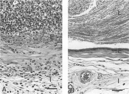
A new animal model of epididymitis due to Chlamydia trachomatis was developed. Adult male Wistar rats were inoculated in the vas deferens with C. trachomatis biovar mouse pneumonitis. After infection, C. trachomatis was recovered from the epididymides for up to 90 days. At day 30, organisms were also reisolated from the testes. Clinical findings included enlargement of infected epididymides and concurrent atrophy of the ipsilateral testes. Histological lesions in the epididymides consisted of pyogranulomatous inflammation, abscesses, and spermatic granulomas. Infection of the testis by C. trachomatis was associated with pyogranulomatous changes. In addition, testicular degeneration, characterized by moderate to severe loss of the germinal epithelium, was noted. Chlamydial antigen was detected within epithelial cells, intratubular macrophages, and macrophages in the stroma of the epididymis by immunoperoxidase staining. This rat model of chlamydial epididymitis appears to clinically and histopathologically mimic the human disease. This model offers the opportunity for further studies on the pathogenesis and sequelae of chlamydial epididymitis.
建立了一种由沙眼衣原体引起的附睾炎新动物模型。成年雄性Wistar大鼠经输精管接种沙眼衣原体生物变种小鼠肺炎衣原体。感染后,在长达90天的时间里,均可从附睾中分离出沙眼衣原体。在第30天,也从睾丸中再次分离出病原体。临床发现包括受感染附睾肿大以及同侧睾丸并发萎缩。附睾的组织学病变包括脓性肉芽肿性炎症、脓肿和精子肉芽肿。沙眼衣原体感染睾丸与脓性肉芽肿性变化有关。此外,还观察到睾丸变性,其特征为生殖上皮中度至重度丧失。通过免疫过氧化物酶染色,在附睾上皮细胞、管内巨噬细胞和间质巨噬细胞中检测到衣原体抗原。这种衣原体附睾炎大鼠模型在临床和组织病理学上似乎模拟了人类疾病。该模型为进一步研究衣原体附睾炎的发病机制和后遗症提供了机会。