Cameron Ivan L, Sun Lu-Zhe, Short Nicholas, Hardman W Elaine, Williams C Douglas
Department of Cellular and Structural Biology, University of Texas Health Science Center, San Antonio, Texas 78229, USA.
Cancer Cell Int. 2005 Jul 26;5:23. doi: 10.1186/1475-2867-5-23.
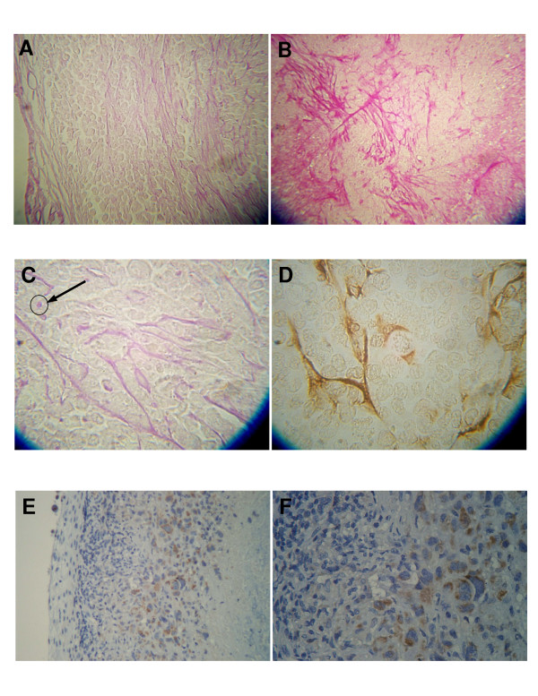
The effects of a rectified semi-sinewave signal (15 mT amplitude, 120 pulses per second, EMF Therapeutics, Inc.) (TEMF) alone and in combination with gamma irradiation (IR) therapy in nude mice bearing a human MDA MB231 breast cancer xenograft were tested. Green fluorescence protein transfected cancer cells were injected into the mammary fat pad of young female mice. Six weeks later, mice were randomly divided into four treatment groups: untreated controls; 10 minute daily TEMF; 200 cGy of IR every other day (total 800 cGy); IR plus daily TEMF. Some mice in each group were euthanized 24 hours after the end of IR. TEMF treatment continued for 3 additional weeks. Tumor sections were stained for: endothelial cells with CD31 and PAS or hypoxia inducible factor 1alpha (HIF).
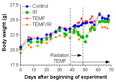
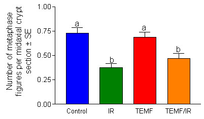
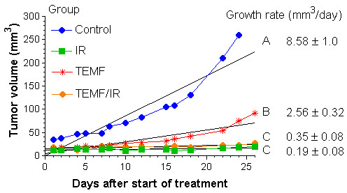
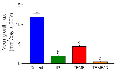
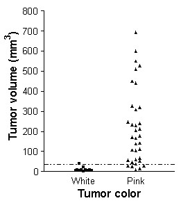
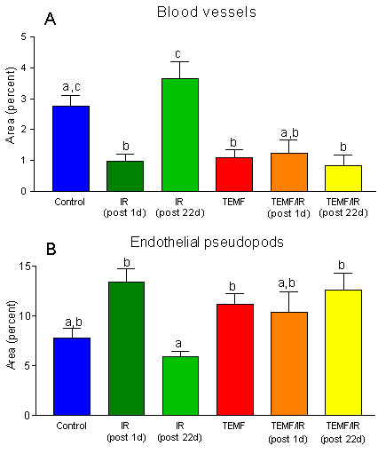
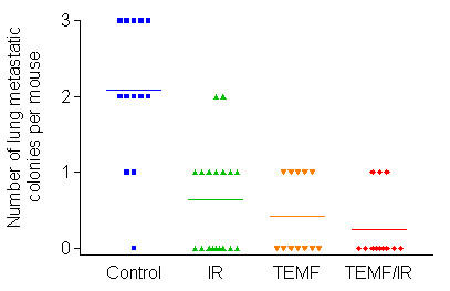
Most tumors <35 mm3 were white but tumors >35 mm3 were pink and had a vascularized capsule. The cortex within 100 microns of the capsule had little vascularization. Blood vessels, capillaries, and endothelial pseudopods were found at >100 microns from the capsule (subcortex). Tumors >35 mm3 treated with IR 24 hours previously or with TEMF had decreased blood vessels in the subcortex and more endothelial pseudopods projecting into hypoxic, HIF positive areas than tumors from the control group. Mice that received either IR or TEMF had significantly fewer lung metastatic sites and slower tumor growth than did untreated mice. No harmful side effects were attributed to TEMF.
TEMF therapy provided a safe means for retarding tumor vascularization, growth and metastasis.
测试了整流半正弦波信号(振幅15 mT,每秒120个脉冲,EMF治疗公司)(TEMF)单独使用以及与γ射线照射(IR)疗法联合使用对携带人MDA MB231乳腺癌异种移植瘤的裸鼠的影响。将绿色荧光蛋白转染的癌细胞注射到年轻雌性小鼠的乳腺脂肪垫中。六周后,将小鼠随机分为四个治疗组:未治疗的对照组;每天进行10分钟的TEMF治疗;每隔一天进行200 cGy的IR照射(总计800 cGy);IR联合每天进行TEMF治疗。每组中的一些小鼠在IR照射结束后24小时实施安乐死。TEMF治疗持续额外的3周。对肿瘤切片进行如下染色:用CD31和PAS或缺氧诱导因子1α(HIF)对内皮细胞进行染色。
大多数体积小于35 mm³的肿瘤为白色,但体积大于35 mm³的肿瘤为粉红色且有血管化的包膜。包膜100微米范围内的皮质几乎没有血管化。在距离包膜(皮质下)超过100微米处发现了血管、毛细血管和内皮伪足。与对照组的肿瘤相比,在24小时前接受IR照射或TEMF治疗的体积大于35 mm³的肿瘤,其皮质下的血管减少且有更多内皮伪足伸向缺氧、HIF阳性区域。接受IR或TEMF治疗的小鼠比未治疗的小鼠肺部转移位点明显更少,肿瘤生长更缓慢。未发现TEMF有有害副作用。
TEMF疗法为延缓肿瘤血管化、生长和转移提供了一种安全的方法。