Department of Cellular and Structural Biology, University of Texas Health Science at San Antonio, 7703 Floyd Curl Drive, San Antonio, Texas 78229 USA.
Research International, Williamsville, New York 14221 USA.
Cancer Cell Int. 2014 Dec 7;14(1):125. doi: 10.1186/s12935-014-0125-5. eCollection 2014.
This study provided additional data on the effects of a therapeutic electromagnetic field (EMF) device on growth and vascularization of murine 16/C mammary adenocarcinoma cells implanted in C3H/HeJ mice.
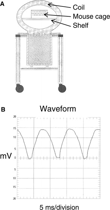
The therapeutic EMF device generated a defined 120 Hz semi sine wave pulse signal of variable intensity. Murine 16/C mammary adenocarcinoma tumor fragments were implanted subcutaneously between the scapulae of syngeneic C3H mice. Once the tumor grew to 100 mm(3), daily EMF treatments were started by placing the cage of mice within the EMF field. Treatment ranged from 10 to 20 milli-Tesla (mT) and was given for 3 to 80 minutes either once or twice a day for 12 days. Tumors were measured and volumes calculated each 3-4 days.
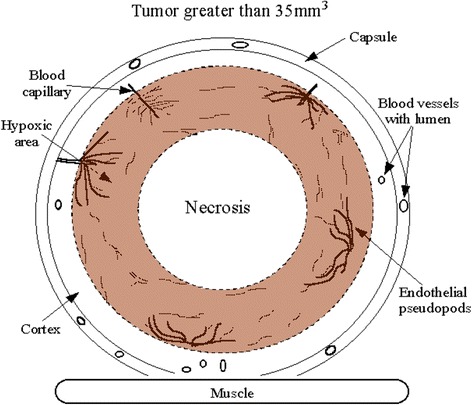
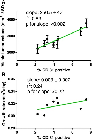
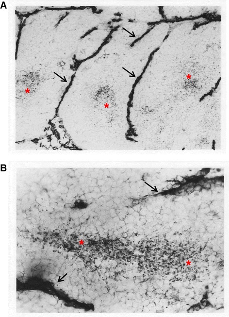
Therapeutic EMF treatment significantly suppressed tumor growth in all 7 EMF treated groups. Exposure to 20mT for 10 minutes twice a day was the most effective tumor growth suppressor. The effect of EMF treatment on extent of tumor vascularization, necrosis and viable area was determined after euthanasia. The EMF reduced the vascular (CD31 immunohistochemically positive) volume fraction and increased the necrotic volume of the tumor. Treatment with 15 mT for 10 min/d gave the maximum anti-angiogenic effect. Lack of a significant correlation between tumor CD 31 positive area and tumor growth rate indicates a mechanism for suppression of tumor growth in addition to suppression of tumor vascularization.
It is proposed that EMF therapy aimed at suppression of tumor growth and vascularization may prove a safe alternative for patients whether they are or are not candidates for conventional cancer therapy.
本研究提供了关于治疗电磁场(EMF)设备对植入 C3H/HeJ 小鼠的 16/C 鼠乳腺腺癌细胞生长和血管生成影响的附加数据。
治疗 EMF 设备产生了可变强度的定义为 120 Hz 的半正弦波脉冲信号。将鼠 16/C 乳腺腺癌肿瘤碎片植入同基因 C3H 小鼠的肩胛骨之间的皮下。一旦肿瘤生长到 100mm3,通过将小鼠笼置于 EMF 场内开始每天进行 EMF 治疗。治疗范围从 10 到 20 毫特斯拉(mT),每天一次或两次,每次 3 到 80 分钟,共 12 天。每隔 3-4 天测量肿瘤并计算体积。
治疗性 EMF 治疗显著抑制了所有 7 个 EMF 治疗组的肿瘤生长。每天两次暴露于 20mT 10 分钟是最有效的肿瘤生长抑制剂。在安乐死后确定 EMF 处理对肿瘤血管生成、坏死和存活区域的影响。EMF 减少了肿瘤的血管(CD31 免疫组化阳性)体积分数并增加了肿瘤的坏死体积。每天 15mT 治疗 10 分钟可获得最大的抗血管生成效果。肿瘤 CD31 阳性面积与肿瘤生长速率之间缺乏显著相关性表明除了抑制肿瘤血管生成外,还有抑制肿瘤生长的机制。
建议针对抑制肿瘤生长和血管生成的 EMF 治疗可能为无论是否为常规癌症治疗候选者的患者提供一种安全的替代方法。