• 文献检索
  • 文档翻译
  • 深度研究
  • 学术资讯
  • Suppr Zotero 插件Zotero 插件
  • 邀请有礼
  • 套餐&价格
  • 历史记录
应用&插件
Suppr Zotero 插件Zotero 插件浏览器插件Mac 客户端Windows 客户端微信小程序
定价
高级版会员购买积分包购买API积分包
服务
文献检索文档翻译深度研究API 文档MCP 服务
关于我们
关于 Suppr公司介绍联系我们用户协议隐私条款
关注我们

Suppr 超能文献

核心技术专利:CN118964589B侵权必究
粤ICP备2023148730 号-1Suppr @ 2026

文献检索

告别复杂PubMed语法,用中文像聊天一样搜索,搜遍4000万医学文献。AI智能推荐,让科研检索更轻松。

立即免费搜索

文件翻译

保留排版,准确专业,支持PDF/Word/PPT等文件格式,支持 12+语言互译。

免费翻译文档

深度研究

AI帮你快速写综述,25分钟生成高质量综述,智能提取关键信息,辅助科研写作。

立即免费体验

脊髓造影、计算机断层扫描和磁共振成像对腰椎管狭窄症跑步机试验预测价值的比较。

Comparison of the predictive value of myelography, computed tomography and MRI on the treadmill test in lumbar spinal stenosis.

作者信息

Moon Eun-Su, Kim Hak-Sun, Park Jin-Oh, Shin Dong-Eun, Ha Jung-Won, Shim Dong-Jun, Kwak Yoon-Hae, Lee Kwang-Il

机构信息

Department of Orthopaedic Surgery, Yongdong Severance Hospital, Yonsei University College of Medicine, Dogok-dong, Kangnam-gu, Seoul 135-720, Korea.

出版信息

Yonsei Med J. 2005 Dec 31;46(6):806-11. doi: 10.3349/ymj.2005.46.6.806.

DOI:10.3349/ymj.2005.46.6.806
PMID:16385657
原文链接:https://pmc.ncbi.nlm.nih.gov/articles/PMC2810595/
Abstract

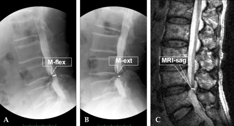
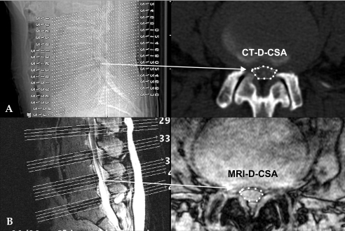
To date, there have been no prospective, objective studies comparing the accuracy of the MRI, myelo-CT and myelography. The purpose of this study is to compare the diagnostic and predictive values of MRIs, myelo-CTs, and myelographies. Myelographies with dynamic motion views, myelo-CTs, MRIs and exercise treadmill tests were performed in 35 cases. The narrowest AP diameter of the dural sac was measured by myelography. At the pathologic level, dural cross-sectional area (D-CSA) was calculated in the MRI and Myelo-CT. The time to the first symptoms (TAF) and the total ambulation time (TAT) were measured during the exercise treadmill test and used as the standard in the comparison of correlation between radiographic parameters and walking capacity. The mean D-CSA by CT was 58.3 mm(2) and 47.6 mm(2) by MRI. All radiographic parameters such as AP diameters and D-CSA have no correlation to TAF or TAT (p > 0.05). Our data showed no statistically significant differences in the correlation of the patients' walking capacity to the severity of stenosis as assessed by myelography, myelo-CT and MRI.

摘要

迄今为止,尚无前瞻性、客观性研究比较MRI、脊髓CT和脊髓造影的准确性。本研究的目的是比较MRI、脊髓CT和脊髓造影的诊断及预测价值。对35例患者进行了动态运动视图脊髓造影、脊髓CT、MRI及运动平板试验。通过脊髓造影测量硬脊膜囊最窄前后径。在病理层面,在MRI和脊髓CT上计算硬脊膜横截面积(D-CSA)。在运动平板试验期间测量首次出现症状的时间(TAF)和总步行时间(TAT),并将其作为比较影像学参数与步行能力之间相关性的标准。CT测量的平均D-CSA为58.3平方毫米,MRI测量的为47.6平方毫米。所有影像学参数,如前后径和D-CSA,与TAF或TAT均无相关性(p>0.05)。我们的数据显示,通过脊髓造影、脊髓CT和MRI评估,患者步行能力与狭窄严重程度之间的相关性无统计学显著差异。

https://cdn.ncbi.nlm.nih.gov/pmc/blobs/8dd9/2810595/6b90a55473f6/ymj-46-806-g002.jpg
https://cdn.ncbi.nlm.nih.gov/pmc/blobs/8dd9/2810595/9b7ed39ca274/ymj-46-806-g001.jpg
https://cdn.ncbi.nlm.nih.gov/pmc/blobs/8dd9/2810595/6b90a55473f6/ymj-46-806-g002.jpg
https://cdn.ncbi.nlm.nih.gov/pmc/blobs/8dd9/2810595/9b7ed39ca274/ymj-46-806-g001.jpg
https://cdn.ncbi.nlm.nih.gov/pmc/blobs/8dd9/2810595/6b90a55473f6/ymj-46-806-g002.jpg

相似文献

1
Comparison of the predictive value of myelography, computed tomography and MRI on the treadmill test in lumbar spinal stenosis.脊髓造影、计算机断层扫描和磁共振成像对腰椎管狭窄症跑步机试验预测价值的比较。
Yonsei Med J. 2005 Dec 31;46(6):806-11. doi: 10.3349/ymj.2005.46.6.806.
2
Axial loading during magnetic resonance imaging in patients with lumbar spinal canal stenosis: does it reproduce the positional change of the dural sac detected by upright myelography?轴向加载在腰椎管狭窄症患者磁共振成像中的应用:它是否能重现直立性椎管造影检测到的硬脊膜囊位置变化?
Spine (Phila Pa 1976). 2012 Jul 15;37(16):E985-92. doi: 10.1097/BRS.0b013e31821038f2.
3
Myelography using flat panel volumetric computed tomography: a comparative study in patients with lumbar spinal stenosis.使用平板容积计算机断层扫描的脊髓造影术:腰椎管狭窄症患者的比较研究
Spine (Phila Pa 1976). 2007 Aug 15;32(18):E523-7. doi: 10.1097/BRS.0b013e3181342673.
4
Improved magnetic resonance myelography of the lumbar spine using image fusion and volumetry.采用图像融合和体绘制技术改善腰椎磁共振脊髓造影。
J Neurosurg Spine. 2014 Feb;20(2):220-6. doi: 10.3171/2013.10.SPINE13119. Epub 2013 Dec 13.
5
[Evaluation of lumbar canal stenosis: decubitus imaging methods versus flexion-extension myelography and surface measurements versus the diameter of the dural sac].腰椎管狭窄症的评估:卧位成像方法与屈伸性脊髓造影对比以及体表测量与硬膜囊直径对比
JBR-BTR. 2000 Apr;83(2):61-7.
6
Comparison of magnetic resonance imaging and computed tomography-myelography for quantitative evaluation of lumbar intracanalar cross-section.磁共振成像与 CT 脊髓造影对腰椎管内横截面积定量评估的比较。
Yonsei Med J. 2011 Jan;52(1):137-44. doi: 10.3349/ymj.2011.52.1.137.
7
Dynamic changes in the dural sac of patients with lumbar canal stenosis evaluated by multidetector-row computed tomography after myelography.脊髓造影后采用多排螺旋计算机断层扫描评估腰椎管狭窄症患者硬脊膜囊的动态变化。
Eur Spine J. 2014 Jan;23(1):74-9. doi: 10.1007/s00586-013-2873-7. Epub 2013 Jul 2.
8
Diagnosis of Lumbar Spinal Stenosis with Functional Myelography.功能性脊髓造影诊断腰椎管狭窄症
J Neurol Surg A Cent Eur Neurosurg. 2018 Jul;79(4):316-322. doi: 10.1055/s-0037-1618563. Epub 2018 Jan 18.
9
Congenital lumbar spinal stenosis: a prospective, control-matched, cohort radiographic analysis.先天性腰椎管狭窄症:一项前瞻性、对照匹配队列影像学分析。
Spine J. 2005 Nov-Dec;5(6):615-22. doi: 10.1016/j.spinee.2005.05.385.
10
The diagnostic value of a treadmill test in predicting lumbar spinal stenosis.平板运动试验对预测腰椎管狭窄症的诊断价值。
Eur Spine J. 2008 May;17(5):686-90. doi: 10.1007/s00586-008-0593-1. Epub 2008 Feb 8.

引用本文的文献

1
Quality of life in lumbar spinal stenosis: Does it correlate with magnetic resonance imaging and spinopelvic parameters?腰椎管狭窄症患者的生活质量:其与磁共振成像及脊柱骨盆参数相关吗?
J Orthop. 2023 Nov 4;47:67-71. doi: 10.1016/j.jor.2023.11.010. eCollection 2024 Jan.
2
Evaluating the Reproducibility of the Walking Test for Intermittent Claudication Associated with Lumbar Spinal Stenosis.评估腰椎管狭窄症相关间歇性跛行步行试验的可重复性。
Asian Spine J. 2022 Jun;16(3):411-418. doi: 10.31616/asj.2020.0505. Epub 2021 May 10.
3
Unilateral Laminectomy by Endoscopy in Central Lumbar Canal Spinal Stenosis: Technical Note and Early Outcomes.

本文引用的文献

1
Imaging lumbar spinal stenosis.
Radiol Clin North Am. 2001 Jan;39(1):31-53, v. doi: 10.1016/s0033-8389(05)70262-1.
2
Degenerative lumbar spinal stenosis.退行性腰椎管狭窄症
J Bone Joint Surg Am. 1998 Jul;80(7):1053-66. doi: 10.2106/00004623-199807000-00015.
3
Use of the exercise treadmill to measure baseline functional status and surgical outcome in patients with severe lumbar spinal stenosis.
Spine (Phila Pa 1976). 1998 Jan 15;23(2):244-8. doi: 10.1097/00007632-199801150-00019.
4
内窥镜下中央椎管狭窄症单侧椎板切除术:技术说明和早期结果。
Spine (Phila Pa 1976). 2020 Jul 15;45(14):E871-E877. doi: 10.1097/BRS.0000000000003478.
4
Indications for prophylactic lumbar decompression at the L3/4 level in patients with L4/5 responsible lumbar spinal canal stenosis.L4/5节段责任性腰椎管狭窄症患者L3/4节段预防性腰椎减压的适应证。
Spine Surg Relat Res. 2017 Nov 27;1(4):191-196. doi: 10.22603/ssrr.1.2016-0032. eCollection 2017.
5
Better Outcomes for Older people with Spinal Trouble (BOOST) Trial: a randomised controlled trial of a combined physical and psychological intervention for older adults with neurogenic claudication, a protocol.老年脊柱问题(BOOST)试验:一项针对伴有神经源性跛行的老年患者的联合物理和心理干预的随机对照试验,方案。
BMJ Open. 2018 Oct 18;8(10):e022205. doi: 10.1136/bmjopen-2018-022205.
6
Postoperative expansion of dural sac cross-sectional area after unilateral laminotomy for bilateral decompression: correlation with clinical symptoms.双侧减压单侧椎板切开术后硬脊膜囊横截面积的术后扩大:与临床症状的相关性
Korean J Spine. 2014 Dec;11(4):227-31. doi: 10.14245/kjs.2014.11.4.227. Epub 2014 Dec 31.
7
Functional and quantitative magnetic resonance myelography of symptomatic stenoses of the lumbar spine.腰椎有症状狭窄的功能和定量磁共振脊髓造影
Neuroradiology. 2014 Dec;56(12):1069-78. doi: 10.1007/s00234-014-1433-0. Epub 2014 Sep 23.
8
Dynamic changes in the dural sac cross-sectional area on axial loaded MR imaging: is there a difference between degenerative spondylolisthesis and spinal stenosis?轴向加载磁共振成像中硬脊膜囊横截面积的动态变化:退行性脊椎滑脱和椎管狭窄之间有区别吗?
AJNR Am J Neuroradiol. 2012 Jun;33(6):1191-7. doi: 10.3174/ajnr.A2920. Epub 2012 Feb 9.
9
Quantification of walking ability in subjects with neurogenic claudication from lumbar spinal stenosis--a comparative study.定量评估腰椎管狭窄症所致神经源性间歇性跛行患者的步行能力——一项对比研究。
Spine J. 2012 Feb;12(2):101-9. doi: 10.1016/j.spinee.2011.12.006. Epub 2011 Dec 29.
10
Myelography in the Age of MRI: Why We Do It, and How We Do It.MRI时代的脊髓造影:我们为何进行以及如何进行。
Radiol Res Pract. 2011;2011:329017. doi: 10.1155/2011/329017. Epub 2011 Mar 6.
Dynamic effects on the lumbar spinal canal: axially loaded CT-myelography and MRI in patients with sciatica and/or neurogenic claudication.对腰椎管的动态影响:坐骨神经痛和/或神经源性间歇性跛行患者的轴向负荷CT脊髓造影和MRI
Spine (Phila Pa 1976). 1997 Dec 15;22(24):2968-76. doi: 10.1097/00007632-199712150-00021.
5
Fluid-fluid levels in a unicameral bone cyst: CT and MR findings.单房性骨囊肿内的液-液平面:CT和MR表现
J Comput Assist Tomogr. 1993 Jan-Feb;17(1):134-6. doi: 10.1097/00004728-199301000-00025.
6
A comparison of computed tomography-myelography, magnetic resonance imaging, and myelography in the diagnosis of herniated nucleus pulposus and spinal stenosis.计算机断层扫描脊髓造影、磁共振成像和脊髓造影在诊断椎间盘突出症和椎管狭窄中的比较。
J Spinal Disord. 1993 Aug;6(4):289-95. doi: 10.1097/00002517-199306040-00002.
7
The predictive value of preoperative myelography in lumbar spinal stenosis.
Spine (Phila Pa 1976). 1994 Jun 15;19(12):1335-8. doi: 10.1097/00007632-199406000-00006.
8
Measurement of exercise tolerance on the treadmill in patients with symptomatic lumbar spinal stenosis: a useful indicator of functional status and surgical outcome.
J Neurosurg. 1995 Jul;83(1):27-30. doi: 10.3171/jns.1995.83.1.0027.
9
Morphometric study of the lumbar spinal canal in the Korean population.韩国人群腰椎管的形态计量学研究。
Spine (Phila Pa 1976). 1995 Aug 1;20(15):1679-84. doi: 10.1097/00007632-199508000-00006.
10
Role of computed tomography and myelography in the diagnosis of central spinal stenosis.计算机断层扫描和脊髓造影在诊断中央型椎管狭窄中的作用。
J Bone Joint Surg Am. 1985 Feb;67(2):240-6.