• 文献检索
  • 文档翻译
  • 深度研究
  • 学术资讯
  • Suppr Zotero 插件Zotero 插件
  • 邀请有礼
  • 套餐&价格
  • 历史记录
应用&插件
Suppr Zotero 插件Zotero 插件浏览器插件Mac 客户端Windows 客户端微信小程序
定价
高级版会员购买积分包购买API积分包
服务
文献检索文档翻译深度研究API 文档MCP 服务
关于我们
关于 Suppr公司介绍联系我们用户协议隐私条款
关注我们

Suppr 超能文献

核心技术专利:CN118964589B侵权必究
粤ICP备2023148730 号-1Suppr @ 2026

文献检索

告别复杂PubMed语法,用中文像聊天一样搜索,搜遍4000万医学文献。AI智能推荐,让科研检索更轻松。

立即免费搜索

文件翻译

保留排版,准确专业,支持PDF/Word/PPT等文件格式,支持 12+语言互译。

免费翻译文档

深度研究

AI帮你快速写综述,25分钟生成高质量综述,智能提取关键信息,辅助科研写作。

立即免费体验

真正组织细胞源性淋巴瘤。所谓真正组织细胞淋巴瘤和恶性组织细胞增多症中不同表型的表达。

Lymphomas of true histiocytic origin. Expression of different phenotypes in so-called true histiocytic lymphoma and malignant histiocytosis.

作者信息

Hsu S M, Ho Y S, Hsu P L

机构信息

Department of Pathology, University of Arkansas for Medical Science, Little Rock.

出版信息

Am J Pathol. 1991 Jun;138(6):1389-404.

PMID:1647137
原文链接:https://pmc.ncbi.nlm.nih.gov/articles/PMC1886401/
Abstract

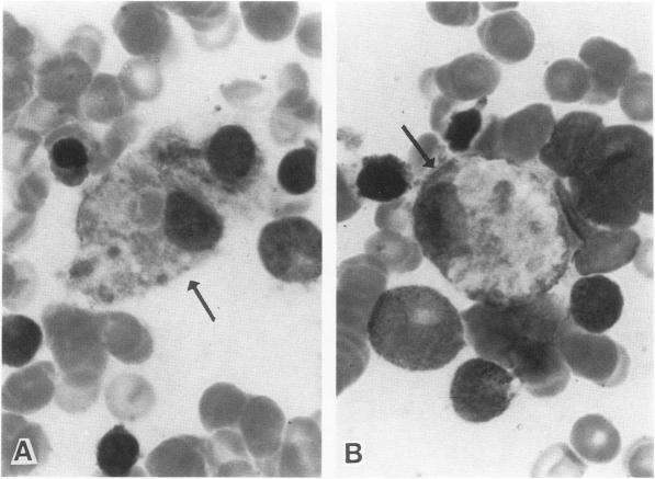
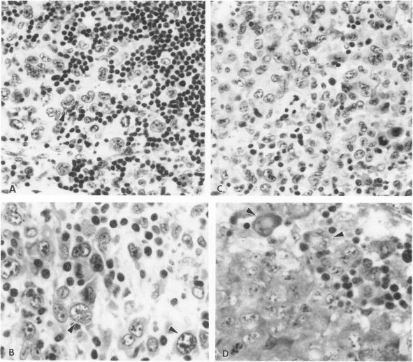
The authors determined the phenotypes of neoplastic cells in true histiocytic lymphoma and malignant histiocytosis by using a large panel of monoclonal antibodies and enzyme histochemistry procedures. Although the phenotypes overlapped slightly, the authors noted a distinct pattern in these tumors. The tumor cells of malignant histiocytosis generally expressed the monocyte markers CD11b, CD11c, CD14, and CD45, especially after induction with phorbol ester. In contrast, the tumor cells of true histiocytic lymphoma exhibited a marker expression very similar to that of Reed-Sternberg cells in Hodgkin's disease. These cells expressed markers CD30, 2H9, and 1A2, but rarely expressed CD11b, CD11c, CD14, or CD45. Regardless of their cytologic features, the tumor cells from both types of histiocytic lymphoma exhibited diffuse nonspecific esterase and acid phosphatase activities, and they expressed histiocyte markers CD15, CD68, LN5, 1E9, and M387 to varying degrees. The tumor cells from both lymphomas did not exhibit T- or B-cell markers, T-cell receptor or immunoglobulin gene rearrangements, or gene translation products, even when they were induced with phorbol ester. The phenotypic expression in these two histiocytic malignancies suggests that they are derived from different types of histiocytes, or from histiocytes in different stages of maturation or differentiation, or from histiocytes that have distinct mechanisms of tumorigenic transformation. The expression of circulating monocyte markers in malignant histiocytosis suggests that this tumor originates in monocytes or free histiocytes, whereas the phenotype of true histiocytic lymphoma is compatible with an origin in fixed histiocytes, which generally are devoid of the monocyte markers CD11b and CD14.

摘要

作者通过使用大量单克隆抗体和酶组织化学方法,确定了真性组织细胞淋巴瘤和恶性组织细胞增多症中肿瘤细胞的表型。尽管这些表型略有重叠,但作者注意到这些肿瘤存在明显的模式。恶性组织细胞增多症的肿瘤细胞通常表达单核细胞标志物CD11b、CD11c、CD14和CD45,尤其是在用佛波酯诱导后。相比之下,真性组织细胞淋巴瘤的肿瘤细胞表现出与霍奇金病里德-斯腾伯格细胞非常相似的标志物表达。这些细胞表达标志物CD30、2H9和1A2,但很少表达CD11b、CD11c、CD14或CD45。无论其细胞学特征如何,两种组织细胞淋巴瘤的肿瘤细胞均表现出弥漫性非特异性酯酶和酸性磷酸酶活性,并且它们不同程度地表达组织细胞标志物CD15、CD68、LN5、1E9和M387。两种淋巴瘤的肿瘤细胞均未表现出T或B细胞标志物、T细胞受体或免疫球蛋白基因重排,或基因翻译产物,即使在用佛波酯诱导时也是如此。这两种组织细胞恶性肿瘤中的表型表达表明,它们源自不同类型的组织细胞,或源自成熟或分化不同阶段的组织细胞,或源自具有不同致瘤转化机制的组织细胞。恶性组织细胞增多症中循环单核细胞标志物的表达表明,这种肿瘤起源于单核细胞或游离组织细胞,而真性组织细胞淋巴瘤的表型与固定组织细胞的起源相符,固定组织细胞通常缺乏单核细胞标志物CD11b和CD14。

https://cdn.ncbi.nlm.nih.gov/pmc/blobs/3a94/1886401/e7cfb24201da/amjpathol00102-0106-a.jpg
https://cdn.ncbi.nlm.nih.gov/pmc/blobs/3a94/1886401/fd5cd9e8104b/amjpathol00102-0099-a.jpg
https://cdn.ncbi.nlm.nih.gov/pmc/blobs/3a94/1886401/7be7d7330300/amjpathol00102-0100-a.jpg
https://cdn.ncbi.nlm.nih.gov/pmc/blobs/3a94/1886401/6fde35d7c1ea/amjpathol00102-0101-a.jpg
https://cdn.ncbi.nlm.nih.gov/pmc/blobs/3a94/1886401/68d53bcbba2d/amjpathol00102-0102-a.jpg
https://cdn.ncbi.nlm.nih.gov/pmc/blobs/3a94/1886401/43126a1aa529/amjpathol00102-0103-a.jpg
https://cdn.ncbi.nlm.nih.gov/pmc/blobs/3a94/1886401/342146bb84f2/amjpathol00102-0104-a.jpg
https://cdn.ncbi.nlm.nih.gov/pmc/blobs/3a94/1886401/97a22624199c/amjpathol00102-0105-a.jpg
https://cdn.ncbi.nlm.nih.gov/pmc/blobs/3a94/1886401/e7cfb24201da/amjpathol00102-0106-a.jpg

相似文献

1
Lymphomas of true histiocytic origin. Expression of different phenotypes in so-called true histiocytic lymphoma and malignant histiocytosis.真正组织细胞源性淋巴瘤。所谓真正组织细胞淋巴瘤和恶性组织细胞增多症中不同表型的表达。
Am J Pathol. 1991 Jun;138(6):1389-404.
2
Phenotypes and phorbol ester-induced differentiation of human histiocytic lymphoma cell lines (U-937 and SU-DHL-1) and Reed-Sternberg cells.人组织细胞淋巴瘤细胞系(U-937和SU-DHL-1)及里德-施特恩伯格细胞的表型和佛波酯诱导的分化
Am J Pathol. 1986 Feb;122(2):223-30.
3
Monoclonal antibodies against SU-DHL-1 cells stain the neoplastic cells in true histiocytic lymphoma, malignant histiocytosis, and Hodgkin's disease.针对SU-DHL-1细胞的单克隆抗体可使真性组织细胞淋巴瘤、恶性组织细胞增多症及霍奇金病中的肿瘤细胞着色。
Blood. 1986 Jul;68(1):213-9.
4
Diagnostic significance of histiocyte-related markers in malignant histiocytosis and true histiocytic lymphoma.组织细胞相关标志物在恶性组织细胞增多症和真性组织细胞淋巴瘤中的诊断意义
Cancer. 1988 Nov 1;62(9):1970-80. doi: 10.1002/1097-0142(19881101)62:9<1970::aid-cncr2820620917>3.0.co;2-v.
5
T-cell/histiocyte-rich large B-cell lymphoma is a disseminated aggressive neoplasm: differential diagnosis from Hodgkin's lymphoma.富于T细胞/组织细胞的大B细胞淋巴瘤是一种播散性侵袭性肿瘤:与霍奇金淋巴瘤的鉴别诊断。
Histopathology. 2002 Sep;41(3):216-29. doi: 10.1046/j.1365-2559.2002.01466.x.
6
Cutaneous histiocytic malignancy. Immunohistochemical re-examination of cases previously diagnosed as cutaneous "histiocytic lymphoma" and "malignant histiocytosis".皮肤组织细胞恶性肿瘤。对先前诊断为皮肤“组织细胞淋巴瘤”和“恶性组织细胞增多症”的病例进行免疫组织化学复查。
J Cutan Pathol. 1993 Apr;20(2):115-20. doi: 10.1111/j.1600-0560.1993.tb00226.x.
7
Cultured Reed-Sternberg cells HDLM-1 and KM-H2 can be induced to become histiocytelike cells. H-RS cells are not derived from lymphocytes.培养的里德-斯腾伯格细胞HDLM-1和KM-H2可被诱导成为组织细胞样细胞。霍奇金里德-斯腾伯格细胞并非来源于淋巴细胞。
Am J Pathol. 1990 Aug;137(2):353-67.
8
High frequency of clonal IG and T-cell receptor gene rearrangements in histiocytic and dendritic cell neoplasms.组织细胞和树突状细胞肿瘤中克隆性免疫球蛋白和T细胞受体基因重排的高频率。
Oncotarget. 2016 Nov 29;7(48):78355-78362. doi: 10.18632/oncotarget.13058.
9
Aberrant expression of T cell and B cell markers in myelocyte/monocyte/histiocyte-derived lymphoma and leukemia cells. Is the infrequent expression of T/B cell markers sufficient to establish a lymphoid origin for Hodgkin's Reed-Sternberg cells?髓细胞/单核细胞/组织细胞来源的淋巴瘤和白血病细胞中T细胞和B细胞标志物的异常表达。霍奇金里德-斯腾伯格细胞中T/B细胞标志物的罕见表达是否足以确定其淋巴样起源?
Am J Pathol. 1989 Jan;134(1):203-12.
10
Frequent immunoglobulin and T-cell receptor gene rearrangements in "histiocytic" neoplasms.“组织细胞性”肿瘤中频繁的免疫球蛋白和T细胞受体基因重排
Am J Pathol. 1985 Dec;121(3):369-73.

引用本文的文献

1
Cerebrospinal fluid soluble CD30 elevation despite suppressive antiretroviral therapy in individuals living with HIV-1.尽管接受了抑制性抗逆转录病毒治疗,但HIV-1感染者的脑脊液可溶性CD30仍升高。
J Virus Erad. 2020 Feb 20;6(1):19-26. doi: 10.1016/S2055-6640(20)30006-6.
2
Malignant histiocytosis of the skin: a case report and review of the literature.皮肤恶性组织细胞增生症:一例报告并文献复习
J Dermatol Case Rep. 2009 Apr 5;3(1):4-7. doi: 10.3315/jdcr.2009.1024.
3
Histiocytoses.组织细胞增多症

本文引用的文献

1
Malignant lymphoma of true histiocytic origin: histiocytic sarcoma. A morphological, ultrastructural, immunological, cytochemical and clinical study of 10 cases.真性组织细胞起源的恶性淋巴瘤:组织细胞肉瘤。10例的形态学、超微结构、免疫学、细胞化学及临床研究
Virchows Arch A Pathol Anat Histol. 1981;391(3):249-65. doi: 10.1007/BF00709158.
2
Production of a monoclonal antibody specific for Hodgkin and Sternberg-Reed cells of Hodgkin's disease and a subset of normal lymphoid cells.制备一种针对霍奇金病的霍奇金细胞和施特恩伯格-里德细胞以及一部分正常淋巴细胞的单克隆抗体。
Nature. 1982 Sep 2;299(5878):65-7. doi: 10.1038/299065a0.
3
Diffuse large cell lymphomas (reticulum cell sarcomas, histiocytic lymphomas). Correlation of morphologic features with functional markers.
Eur J Pediatr. 1995 Aug;154(8):600-9. doi: 10.1007/BF02079059.
弥漫性大细胞淋巴瘤(网状细胞肉瘤、组织细胞淋巴瘤)。形态学特征与功能标志物的相关性。
Cancer. 1980 Sep 15;46(6):1428-41. doi: 10.1002/1097-0142(19800915)46:6<1428::aid-cncr2820460624>3.0.co;2-r.
4
True histiocytic lymphoma.真性组织细胞淋巴瘤
Arch Dermatol. 1980 Aug;116(8):915-8.
5
A monoclonal antibody reactive with human peripheral blood monocytes.一种与人外周血单核细胞反应的单克隆抗体。
J Immunol. 1980 Apr;124(4):1943-8.
6
A monoclonal antibody (anti-Tac) reactive with activated and functionally mature human T cells. I. Production of anti-Tac monoclonal antibody and distribution of Tac (+) cells.一种与活化且功能成熟的人T细胞反应的单克隆抗体(抗Tac)。I. 抗Tac单克隆抗体的产生及Tac(+)细胞的分布。
J Immunol. 1981 Apr;126(4):1393-7.
7
Malignant histiocytosis. Ultrastructural and immunocytochemical characterization.恶性组织细胞增多症。超微结构及免疫细胞化学特征
Arch Pathol Lab Med. 1980 Jan;104(1):26-9.
8
Acute leukaemia with mixed lymphoid and myeloid phenotype.具有混合淋巴样和髓样表型的急性白血病。
Br J Haematol. 1984 Jan;56(1):121-30. doi: 10.1111/j.1365-2141.1984.tb01277.x.
9
Acute leukaemia with mixed lymphoid and myeloid phenotype.具有混合淋巴样和髓样表型的急性白血病。
Br J Haematol. 1984 Sep;58(1):203-5. doi: 10.1111/j.1365-2141.1984.tb06075.x.
10
Phenotypic expression of B-lymphocytes. 1. Identification with monoclonal antibodies in normal lymphoid tissues.B淋巴细胞的表型表达。1. 在正常淋巴组织中用单克隆抗体进行鉴定。
Am J Pathol. 1984 Mar;114(3):387-95.