Department of Hazardous Microorganisms, Walter Reed Army Institute of Research, Washington, D.C. 20012.
Infect Immun. 1974 Nov;10(5):1152-5. doi: 10.1128/iai.10.5.1152-1155.1974.
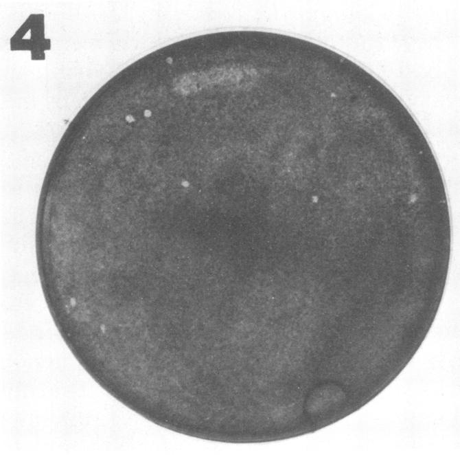
Mammalian cells particularly suitable for the study of specialized aspects of rickettsial biology were tested for their ability to support plaque formation by Rickettsia conori. The detection of plaques was substantially influenced by the combination of growth medium and cell type used. Large plaques (2.0 to 3.0 mm in diameter) occurred by 8 days postinfection in WI-38 and DBS-FRhL-2 cells supported by medium 199. Smaller plaques (0.5 to 1.0 mm in diameter) were seen in L-929 and HeLa cells at 8 to 11 days postinfection and were more discernible in cells supported with Eagle minimal essential medium. Chicken embryo cells maintained in Dulbecco's modified Eagle medium exhibited large spherical plaques with a diameter of approximately 1.5 mm by 8 days postinfection.
检测了特别适合研究立克次体生物学特殊方面的哺乳动物细胞,以确定它们支持考德里氏立克次体形成斑的能力。噬菌斑的检测受到所使用的生长培养基和细胞类型组合的显著影响。在感染后 8 天,WI-38 和 DBS-FRhL-2 细胞在 199 号培养基中支持下形成大的噬菌斑(直径 2.0 至 3.0 毫米)。在 L-929 和 HeLa 细胞中,在感染后 8 至 11 天出现较小的噬菌斑(直径 0.5 至 1.0 毫米),在使用 Eagle 最低必需培养基支持的细胞中更容易识别。在改良的 Eagle 培养基中维持的鸡胚细胞在感染后 8 天形成直径约 1.5 毫米的大球形噬菌斑。