Ferko Michael C, Patterson Brian W, Butler Peter J
Department of Bioengineering, The Pennsylvania State University, University Park, Pennsylvania 16802, USA.
Microsc Res Tech. 2006 Aug;69(8):648-55. doi: 10.1002/jemt.20332.
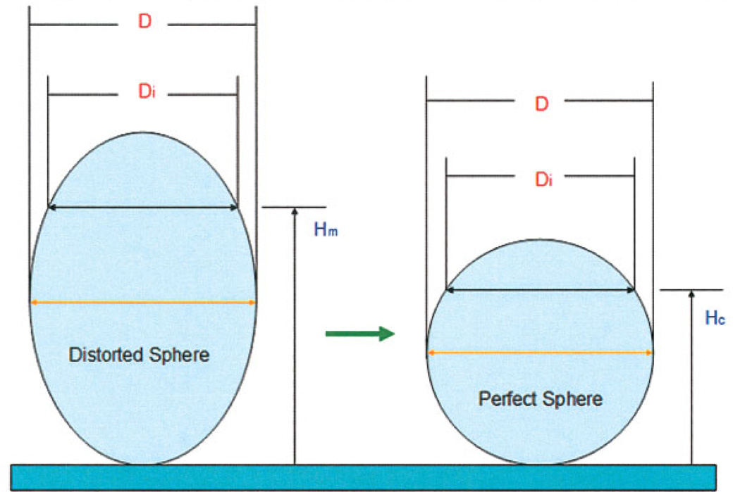
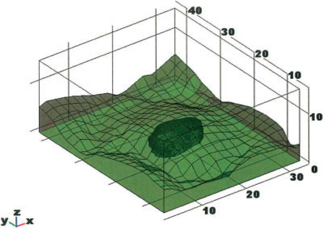
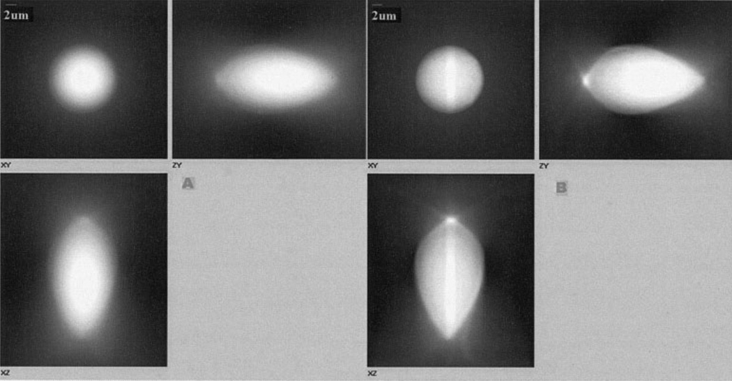
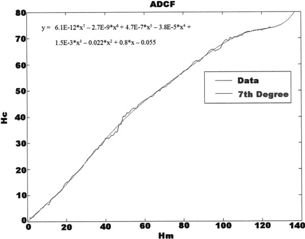
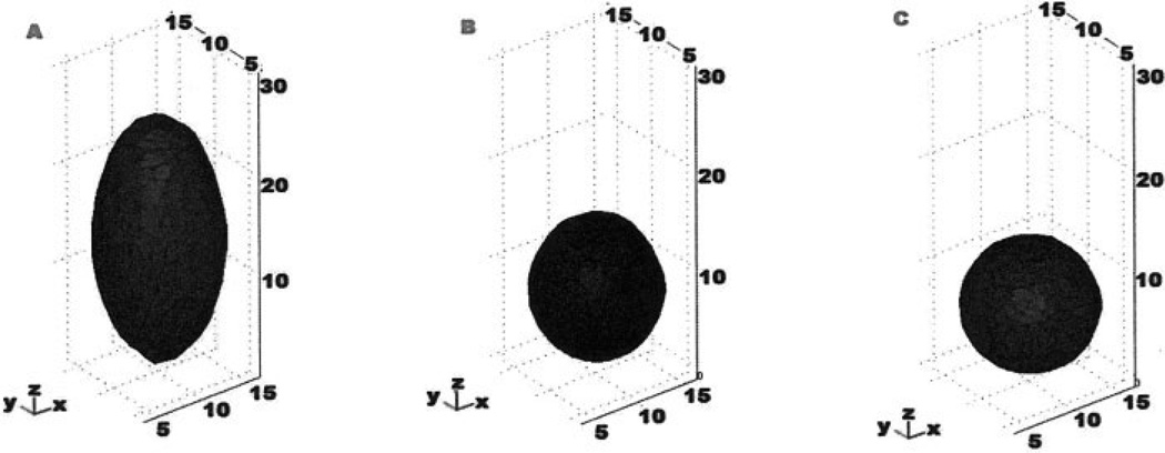
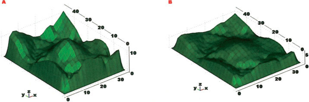
Optical-sectioning, digital fluorescence microscopy provides images representing temporally- and spatially-resolved molecular-scale details of the substructures of living cells. To render such images into solid models for further computational analyses, we have developed an integrated system of image acquisition, processing, and rendering, which includes a new empirical technique to correct for axial distortions inherent in fluorescence microscopy due to refractive index mismatches between microscope objective immersion medium, coverslip glass, and water. This system takes advantage of the capabilities of ultra-high numerical aperture objectives (e.g. total internal reflection fluorescence microscopy) and enables faithful three-dimensional rendering of living cells into solid models amenable to further computational analysis. An example of solid modeling of bovine aortic endothelial cells and their nuclei is presented. Since many cellular level events are temporally and spatially confined, such integrated image acquisition, processing, rendering, and computational analysis, will enable, in silico, the generation of new computational models for cell mechanics and signaling.
光学切片数字荧光显微镜能够提供代表活细胞亚结构分子尺度细节的时空分辨图像。为了将这些图像转化为实体模型以进行进一步的计算分析,我们开发了一个图像采集、处理和渲染的集成系统,其中包括一种新的经验技术,用于校正由于显微镜物镜浸没介质、盖玻片和水之间的折射率不匹配而导致的荧光显微镜固有的轴向畸变。该系统利用了超高数值孔径物镜的功能(例如全内反射荧光显微镜),能够将活细胞忠实地三维渲染成适合进一步计算分析的实体模型。文中给出了牛主动脉内皮细胞及其细胞核的实体建模示例。由于许多细胞水平的事件在时间和空间上受到限制,这种集成的图像采集、处理、渲染和计算分析将能够在计算机上生成用于细胞力学和信号传导的新计算模型。