Koopmans Steven A, Terwee Thom, Glasser Adrian, Wendt Mark, Vilupuru Abhiram S, van Kooten Theo G, Norrby Sverker, Haitjema Henk J, Kooijman Aart C
Department of Ophthalmology, University Medical Center Groningen, University of Groningen, The Netherlands.
Invest Ophthalmol Vis Sci. 2006 Jul;47(7):2976-84. doi: 10.1167/iovs.05-1346.
Accommodation can be restored to presbyopic human eyes by refilling the capsular bag with a soft polymer. This study was conducted to test whether accommodation, measurable as changes in optical refraction, can be restored with a newly developed refilling polymer in a rhesus monkey model. A specific intra- and postoperative treatment protocol was used to minimize postoperative inflammation and to delay capsular opacification.
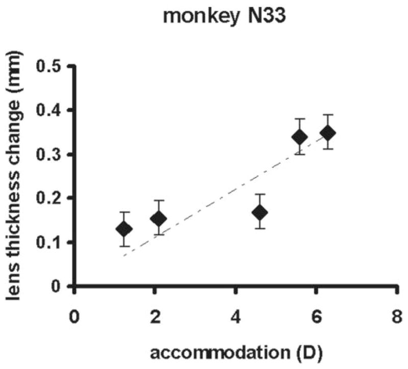
Nine adolescent rhesus monkeys underwent refilling of the lens capsular bag with a polymer. In the first four monkeys (group A) the surgical procedure was followed by two weekly subconjunctival injections of corticosteroids. In a second group of five monkeys (group B) a treatment intended to delay the development of capsular opacification was applied during the surgery, and, in the postoperative period, eye drops and two subconjunctival injections of corticosteroids were applied. Accommodation was stimulated with carbachol iontophoresis or pilocarpine and was measured with a Hartinger refractometer at regular times during a follow-up period of 37 weeks in five monkeys. In one monkey, lens thickness changes were measured with A-scan ultrasound.
In group A, refraction measurement was possible in one monkey. In the three other animals in group A, postoperative inflammation and capsular opacification prevented refraction measurements. In group B, the maximum accommodative amplitude of the surgically treated eyes was 6.3 D. In three monkeys the accommodative amplitude decreased to almost 0 D after 37 weeks. In the two other monkeys, the accommodative amplitude remained stable at +/-4 D during the follow-up period. In group B, capsular opacification developed in the postoperative period, but refraction measurements could still be performed during the whole follow-up period of 37 weeks.
A certain level of accommodation can be restored after lens refilling in adolescent rhesus monkeys. During the follow-up period refraction measurements were possible in all five monkeys that underwent the treatment designed to prevent inflammation and capsular opacification.
通过向晶状体囊袋内重新填充一种软性聚合物,可恢复老视人眼的调节功能。本研究旨在测试在恒河猴模型中,使用一种新开发的填充聚合物是否能恢复可通过眼屈光变化测量的调节功能。采用了特定的术中和术后治疗方案,以尽量减少术后炎症并延缓晶状体后囊混浊。
对9只青春期恒河猴的晶状体囊袋进行聚合物填充。在前4只猴子(A组)中,手术后每周进行两次结膜下注射皮质类固醇。在第二组5只猴子(B组)中,手术期间采用了旨在延缓晶状体后囊混浊发展的治疗方法,术后使用眼药水并进行两次结膜下注射皮质类固醇。通过卡巴胆碱离子导入或毛果芸香碱刺激调节功能,并在5只猴子37周的随访期内定期用Hartinger验光仪进行测量。在1只猴子中,用A超测量晶状体厚度变化。
在A组中,仅1只猴子能够进行屈光测量。A组的其他3只动物术后炎症和晶状体后囊混浊妨碍了屈光测量。在B组中,手术治疗眼的最大调节幅度为6.3D。3只猴子在37周后调节幅度降至几乎为0D。另外2只猴子在随访期内调节幅度保持在±4D稳定。在B组中,术后出现了晶状体后囊混浊,但在37周的整个随访期内仍可进行屈光测量。
青春期恒河猴晶状体填充后可恢复一定程度的调节功能。在接受旨在预防炎症和晶状体后囊混浊治疗的所有5只猴子的随访期内,均可进行屈光测量。