Gu Qiang, Sivanandam Thamil Mani, Kim Caroline Aehyun
Department of Neurobiology and Anatomy, Wake Forest University School of Medicine, Medical Center Boulevard, Winston-Salem, North Carolina 27157, USA.
Proteome Sci. 2006 Oct 11;4:21. doi: 10.1186/1477-5956-4-21.
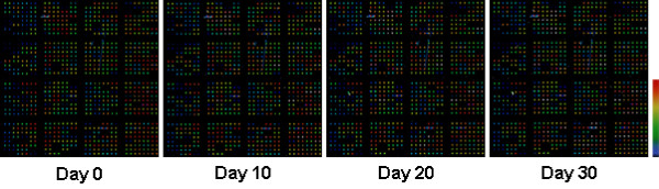
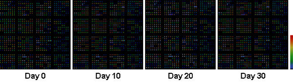
The antibody microarray technique is a newly emerging proteomics tool for differential protein expression analyses that uses fluorescent dyes Cy 3 and Cy 5. Environmental factors, such as light exposure, can affect the signal intensity of fluorescent dyes on microarray slides thus, it is logical to scan microarray slides immediately after the final wash and drying processes. However, no research data are available concerning time-dependent changes of fluorescent signals on antibody microarray slides to this date. In the present study, microarray slides were preserved at -20 degrees C after regular microarray experiments and were rescanned at day 10, 20 and 30 to evaluate change in signal intensity.
Fluorescent intensities of microarray spots were detected using a confocal laser scanner after the experiment at day 0, and re-examined at day 10, 20 and 30, respectively. Fluorescent intensities of rescanned microarray spots did not show significant changes when compared with those scanned immediately after standard microarray experiments.
Microarray slides can be preserved and rescanned multiple times using a confocal laser scanner over a period of days or weeks.
抗体微阵列技术是一种新兴的蛋白质组学工具,用于差异蛋白质表达分析,它使用荧光染料Cy 3和Cy 5。环境因素,如光照,会影响微阵列载玻片上荧光染料的信号强度,因此,在最后洗涤和干燥过程后立即扫描微阵列载玻片是合理的。然而,迄今为止,尚无关于抗体微阵列载玻片上荧光信号随时间变化的研究数据。在本研究中,微阵列载玻片在常规微阵列实验后保存在-20℃,并在第10、20和30天重新扫描以评估信号强度的变化。
在第0天实验后使用共聚焦激光扫描仪检测微阵列斑点的荧光强度,并分别在第10、20和30天重新检查。与标准微阵列实验后立即扫描的结果相比,重新扫描的微阵列斑点的荧光强度没有显示出显著变化。
微阵列载玻片可以在几天或几周的时间内使用共聚焦激光扫描仪进行保存和多次重新扫描。