Gubbels Jennifer A A, Belisle Jennifer, Onda Masanori, Rancourt Claudine, Migneault Martine, Ho Mitchell, Bera Tapan K, Connor Joseph, Sathyanarayana Bangalore K, Lee Byungkook, Pastan Ira, Patankar Manish S
Department of Obstetrics and Gynecology, University of Wisconsin-Madison, Madison, USA.
Mol Cancer. 2006 Oct 26;5(1):50. doi: 10.1186/1476-4598-5-50.
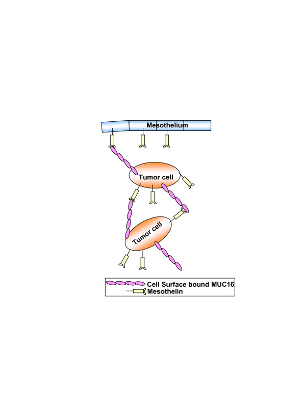
The mucin MUC16 and the glycosylphosphatidylinositol anchored glycoprotein mesothelin likely facilitate the peritoneal metastasis of ovarian tumors. The biochemical basis and the kinetics of the binding between these two glycoproteins are not clearly understood. Here we have addressed this deficit and provide further evidence supporting the role of the MUC16-mesothelin interaction in facilitating cell-cell binding under conditions that mimic the peritoneal environment.
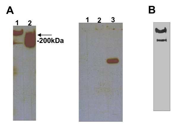
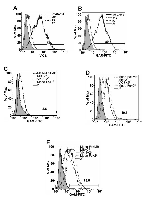
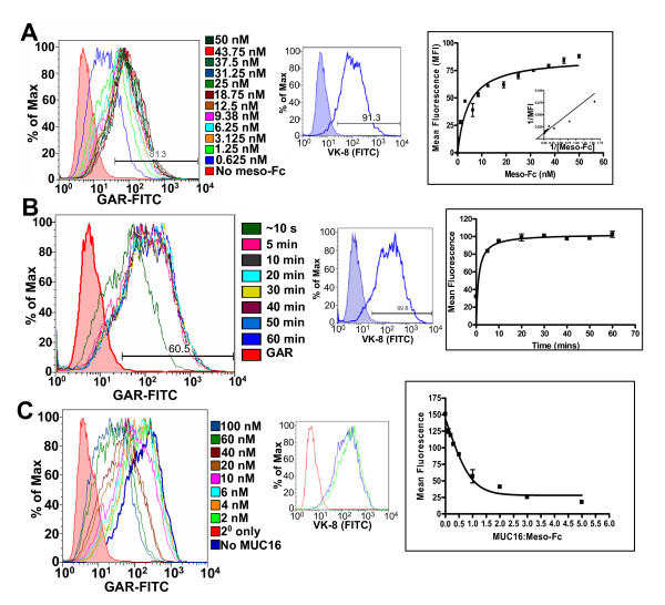
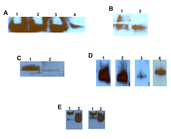
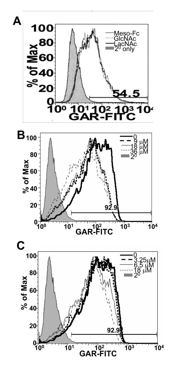
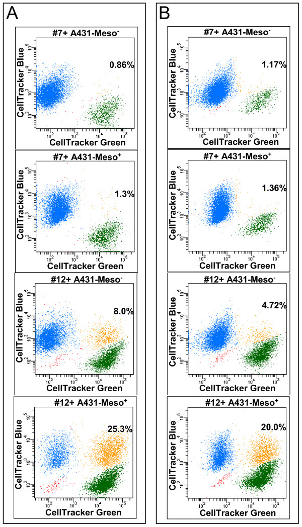
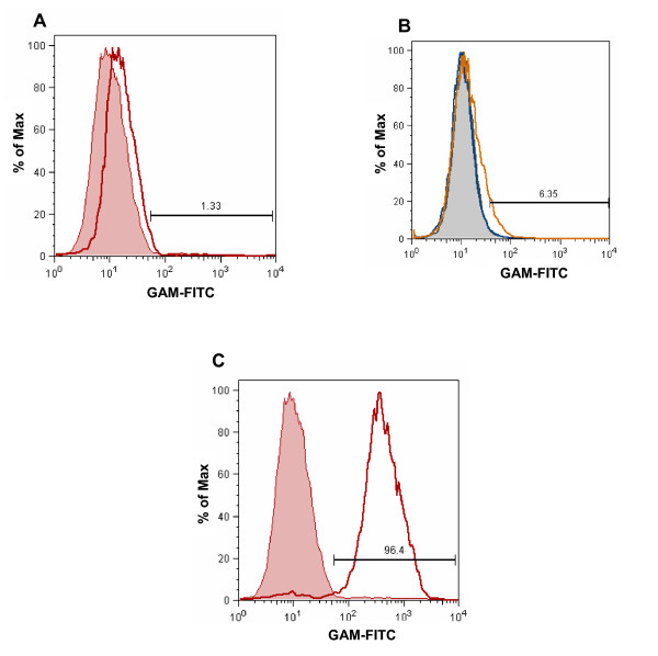
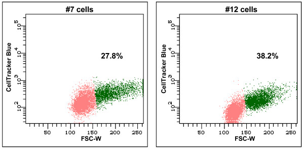
In this study we utilize recombinant-Fc tagged human mesothelin to measure the binding kinetics of this glycoprotein to MUC16 expressed on the ovarian tumor cell line OVCAR-3. OVCAR-3 derived sublines that did not express MUC16 showed no affinity for mesothelin. In a flow cytometry-based assay mesothelin binds with very high affinity to the MUC16 on the OVCAR-3 cells with an apparent Kd of 5-10 nM. Maximum interaction occurs within 5 mins of incubation of the recombinant mesothelin with the OVCAR-3 cells and significant binding is observed even after 10 sec. A five-fold molar excess of soluble MUC16 was unable to completely inhibit the binding of mesothelin to the OVCAR-3 cells. Oxidation of the MUC16 glycans, removal of its N-linked oligosaccharides, and treatment of the mucin with wheat germ agglutinin and erythroagglutinating phytohemagglutinin abrogates its binding to mesothelin. These observations suggest that at least a subset of the MUC16-asscociated N-glycans is required for binding to mesothelin. We also demonstrate that MUC16 positive ovarian tumor cells exhibit increased adherence to A431 cells transfected with mesothelin (A431-Meso+). Only minimal adhesion is observed between MUC16 knockdown cells and A431-Meso+ cells. The binding between the MUC16 expressing ovarian tumor cells and the A431-Meso+ cells occurs even in the presence of ascites from patients with ovarian cancer.
The strong binding kinetics of the mesothelin-MUC16 interaction and the cell adhesion between ovarian tumor cells and A431-Meso+ even in the presence of peritoneal fluid strongly support the importance of these two glycoproteins in the peritoneal metastasis of ovarian tumors. The demonstration that N-linked glycans are essential for mediating mesothlein-MUC16 binding may lead to novel therapeutic targets to control the spread of ovarian carcinoma.
黏蛋白MUC16和糖基磷脂酰肌醇锚定糖蛋白间皮素可能促进卵巢肿瘤的腹膜转移。这两种糖蛋白之间结合的生化基础和动力学尚不清楚。在此,我们解决了这一不足,并提供了进一步的证据,支持MUC16-间皮素相互作用在模拟腹膜环境的条件下促进细胞间结合中的作用。
在本研究中,我们利用重组Fc标签的人源间皮素来测量这种糖蛋白与卵巢肿瘤细胞系OVCAR-3上表达的MUC16的结合动力学。不表达MUC16的OVCAR-3衍生亚系对间皮素无亲和力。在基于流式细胞术的检测中,间皮素以非常高的亲和力与OVCAR-3细胞上的MUC16结合,表观解离常数(Kd)为5-10 nM。重组间皮素与OVCAR-3细胞孵育5分钟内发生最大相互作用,即使在10秒后也能观察到显著结合。五倍摩尔过量的可溶性MUC16不能完全抑制间皮素与OVCAR-3细胞的结合。MUC16聚糖的氧化、其N-连接寡糖的去除以及用麦胚凝集素和红细胞凝集植物血凝素处理黏蛋白可消除其与间皮素的结合。这些观察结果表明,至少一部分与MUC16相关的N-聚糖是与间皮素结合所必需的。我们还证明,MUC16阳性的卵巢肿瘤细胞对转染了间皮素的A431细胞(A431-Meso+)的黏附增加。在MUC16敲低细胞和A431-Meso+细胞之间仅观察到最小程度的黏附。表达MUC16的卵巢肿瘤细胞与A431-Meso+细胞之间的结合即使在存在卵巢癌患者腹水的情况下也会发生。
间皮素-MUC16相互作用的强结合动力学以及卵巢肿瘤细胞与A431-Meso+细胞之间即使在存在腹膜液的情况下的细胞黏附,有力地支持了这两种糖蛋白在卵巢肿瘤腹膜转移中的重要性。N-连接聚糖对于介导间皮素-MUC16结合至关重要的证明可能会导致控制卵巢癌扩散的新治疗靶点。