Stella Salvatore L, Hu Wanda D, Vila Alejandro, Brecha Nicholas C
Department of Neurobiology, David Geffen School of Medicine, University of California, Los Angeles, California, USA.
J Neurosci Res. 2007 Apr;85(5):1126-37. doi: 10.1002/jnr.21210.
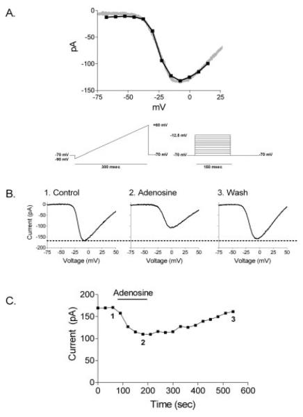
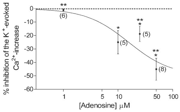
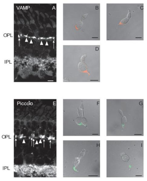
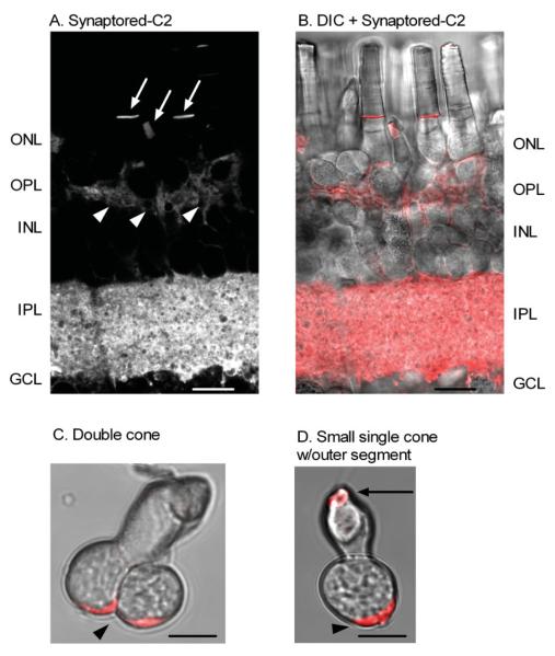
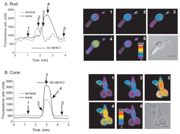
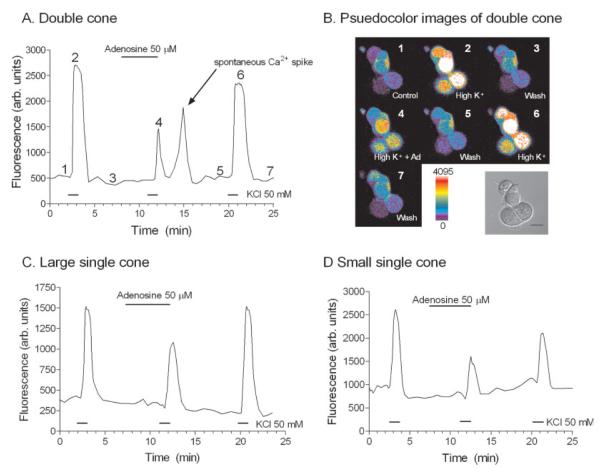
Endogenous adenosine has already been shown to inhibit transmitter release from the rod synapse by suppressing Ca(2+) influx through voltage-gated Ca(2+) channels. However, it is not clear how adenosine modulates the cone synapse. Cone photoreceptors, like rod photoreceptors, also possess L-type Ca(2+) channels that regulate the release of L-glutamate. To assess the impact of adenosine on Ca(2+) influx though voltage-gated Ca(2+) channels in cone terminals, whole-cell perforated-patch clamp recording and Ca(2+) imaging with fluo-4 were used on isolated cones and salamander retinal slices. Synaptic markers (VAMP and piccolo) and activity-dependent dye labeling revealed that tiger salamander cone terminals contain a broad, vesicle-filled cytoplasmic extension at the base of the somatic compartment, which is unlike rod terminals that contain one or more thin axons, each terminating in a large bulbous synaptic terminal. The spatiotemporal Ca(2+) responses of the cone terminals do not differ significantly from the Ca(2+) responses of the soma or inner segment like that observed in rods. Whole-cell recording of cone I(Ca) and Ca(2+) imaging of synaptic terminals in cones demonstrate that adenosine inhibited both I(Ca) and the depolarization-evoked Ca(2+) increase in cone terminals in a dose-dependent manner from 1 to 50 muM. These results indicate that, as in rods, adenosine's ability to suppress voltage-dependent Ca(2+) channels at the cone synapse will limit the amount of L-glutamate released. Therefore, adenosine has an inhibitory effect on L-glutamate release at the first synapse, which likely favors elevated adenosine levels in the dark or during dark-adapted conditions.
内源性腺苷已被证明可通过抑制电压门控钙通道的钙内流来抑制视杆突触的递质释放。然而,腺苷如何调节视锥突触尚不清楚。视锥光感受器与视杆光感受器一样,也拥有调节L-谷氨酸释放的L型钙通道。为了评估腺苷对视锥终末电压门控钙通道钙内流的影响,对分离的视锥细胞和蝾螈视网膜切片进行了全细胞膜片钳穿孔记录和用Fluo-4进行的钙成像。突触标记物(VAMP和 piccolo)以及活性依赖的染料标记显示,虎蝾螈视锥终末在体细胞区基部含有一个宽阔的、充满囊泡的细胞质延伸部分,这与视杆终末不同,视杆终末含有一条或多条细轴突,每条轴突在一个大的球状突触终末终止。视锥终末的时空钙反应与视杆细胞中观察到的体细胞或内段的钙反应没有显著差异。视锥细胞I(Ca)的全细胞记录和视锥突触终末的钙成像表明,腺苷以1至50 μM的剂量依赖性方式抑制视锥终末的I(Ca)和去极化诱发的钙增加。这些结果表明,与视杆细胞一样,腺苷在视锥突触处抑制电压依赖性钙通道的能力将限制L-谷氨酸的释放量。因此,腺苷对第一个突触处的L-谷氨酸释放具有抑制作用,这可能有利于在黑暗或暗适应条件下腺苷水平的升高。