Whitwell Jennifer L, Jack Clifford R, Parisi Joseph E, Knopman David S, Boeve Bradley F, Petersen Ronald C, Ferman Tanis J, Dickson Dennis W, Josephs Keith A
Department of Radiology, Mayo Clinic Rochester, Rochester, MN 55905, USA.
Brain. 2007 Apr;130(Pt 4):1148-58. doi: 10.1093/brain/awm021. Epub 2007 Mar 8.
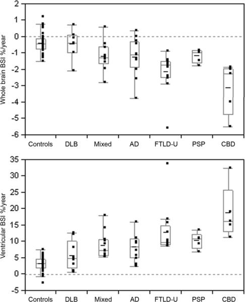
Neurodegenerative disorders are pathologically characterized by the deposition of abnormal proteins in the brain. It is likely that future treatment trials will target the underlying protein biochemistry and it is therefore increasingly important to be able to distinguish between different pathologies during life. The aim of this study was to determine whether rates of brain atrophy differ in neurodegenerative dementias that vary by pathological diagnoses and characteristic protein biochemistry. Fifty-six autopsied subjects were identified with a clinical diagnosis of dementia and two serial head MRI. Subjects were subdivided based on pathological diagnoses into Alzheimer's disease, dementia with Lewy bodies (DLB), mixed Alzheimer's disease/DLB, frontotemporal lobar degeneration with ubiquitin-only-immunoreactive changes (FTLD-U), corticobasal degeneration (CBD) and progressive supranuclear palsy (PSP). Twenty-five controls were matched by age, gender and scan interval, to the study cohort. The boundary-shift integral was used to calculate change over time in whole brain (BBSI) and ventricular volume (VBSI). All BSI results were annualized by adjusting for scan interval. The rates of whole brain atrophy and ventricular expansion were significantly increased compared to controls in the Alzheimer's disease, mixed Alzheimer's disease/DLB, FTLD-U, CBD and PSP groups. However, atrophy rates in the DLB group were not significantly different from control rates of atrophy. The largest rates of atrophy were observed in the CBD group which had a BBSI of 2.3% and VBSI of 16.2%. The CBD group had significantly greater rates of BBSI and VBSI than the DLB, mixed Alzheimer's disease/DLB, Alzheimer's disease and PSP groups, with a similar trend observed when compared to the FTLD-U group. The FTLD-U group showed the next largest rates with a BBSI of 1.7% and VBSI of 9.6% which were both significantly greater than the DLB group. There was no significant difference in the rates of atrophy between the Alzheimer's disease, mixed Alzheimer's disease/DLB and PSP groups, which all showed similar rates of atrophy; BBSI of 1.1, 1.3 and 1.0% and VBSI of 8.3, 7.2 and 10.9%, respectively. Rates of atrophy therefore differ according to the pathological diagnoses and underlying protein biochemistry. While rates are unlikely to be useful in differentiating Alzheimer's disease from cases with mixed Alzheimer's disease/DLB pathology, they demonstrate important pathophysiological differences between DLB and those with mixed Alzheimer's disease/DLB and Alzheimer's disease pathology, and between those with CBD and PSP pathology.
神经退行性疾病的病理特征是大脑中异常蛋白质的沉积。未来的治疗试验可能会针对潜在的蛋白质生物化学,因此在生命过程中能够区分不同的病理情况变得越来越重要。本研究的目的是确定脑萎缩率在因病理诊断和特征性蛋白质生物化学而异的神经退行性痴呆中是否存在差异。56名经尸检的受试者被临床诊断为痴呆,并进行了两次连续的头部MRI检查。受试者根据病理诊断被细分为阿尔茨海默病、路易体痴呆(DLB)、阿尔茨海默病/DLB混合型、仅伴有泛素免疫反应性改变的额颞叶变性(FTLD-U)、皮质基底节变性(CBD)和进行性核上性麻痹(PSP)。25名对照者在年龄、性别和扫描间隔方面与研究队列相匹配。边界位移积分用于计算全脑(BBSI)和脑室容积(VBSI)随时间的变化。所有BSI结果均通过调整扫描间隔进行年化处理。与对照组相比,阿尔茨海默病、阿尔茨海默病/DLB混合型、FTLD-U、CBD和PSP组的全脑萎缩率和脑室扩张率显著增加。然而,DLB组的萎缩率与对照组的萎缩率无显著差异。萎缩率最高的是CBD组,其BBSI为2.3%,VBSI为16.2%。CBD组的BBSI和VBSI率显著高于DLB组、阿尔茨海默病/DLB混合型、阿尔茨海默病组和PSP组,与FTLD-U组相比也观察到类似趋势。FTLD-U组的萎缩率次之,BBSI为1.7%,VBSI为9.6%,均显著高于DLB组。阿尔茨海默病组、阿尔茨海默病/DLB混合型和PSP组的萎缩率无显著差异,它们的萎缩率相似;BBSI分别为1.1%、1.3%和1.0%,VBSI分别为8.3%、7.2%和10.9%。因此,萎缩率因病理诊断和潜在的蛋白质生物化学而异。虽然萎缩率不太可能用于区分阿尔茨海默病与阿尔茨海默病/DLB混合型病例,但它们显示了DLB与阿尔茨海默病/DLB混合型和阿尔茨海默病病例之间,以及CBD与PSP病例之间重要的病理生理差异。