Indian Institute of Science Education and Research (IISER) Berhampur, Berhampur, India.
Transl Psychiatry. 2024 Sep 27;14(1):389. doi: 10.1038/s41398-024-03097-2.
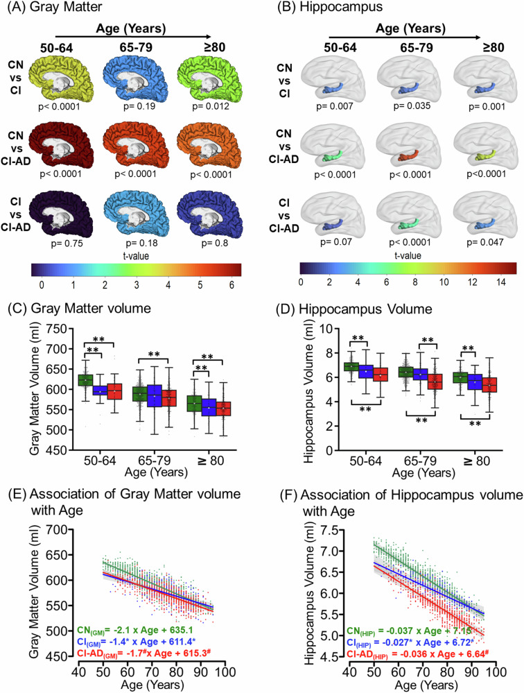
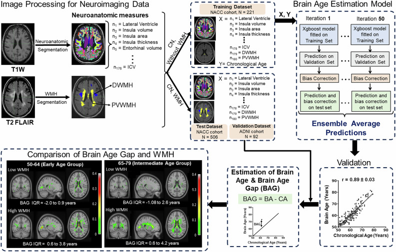
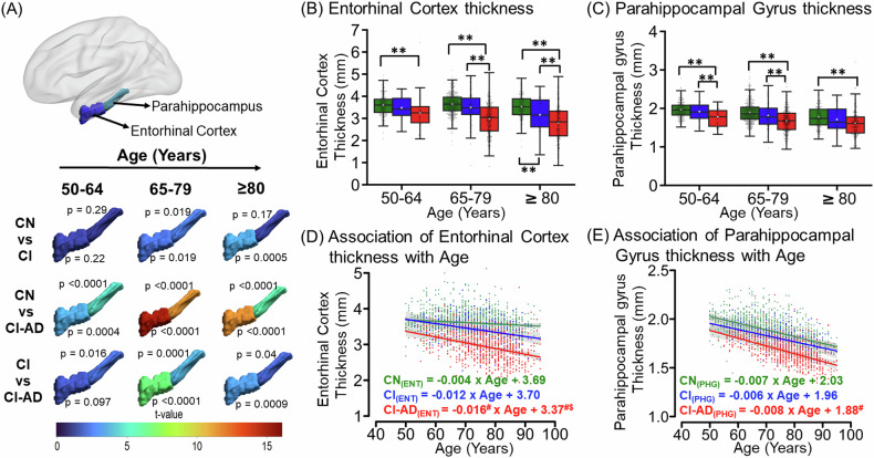
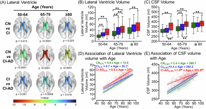
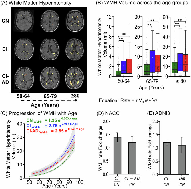
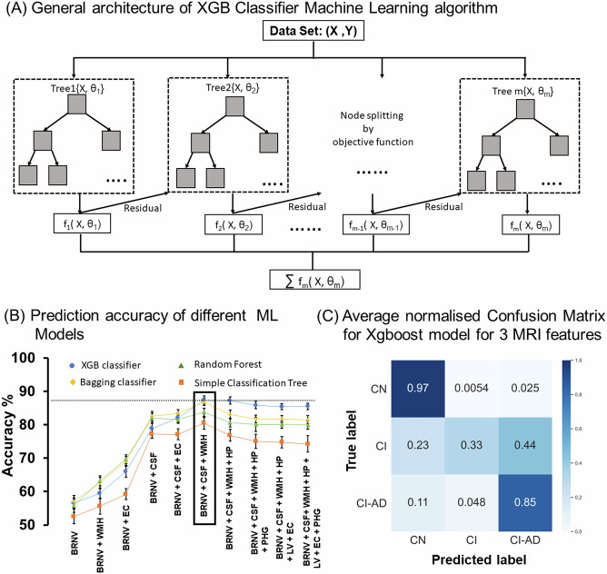
Even among the subjects classified as cognitively normal, there exists a subset of individuals at a given chronological age (CA) who harbor white matter hyperintensity (WMH) while another subset presents with low or undetectable WMH. Here, we conducted a comprehensive MRI segmentation of neuroanatomic structures along with WMH quantification in groups of cognitively normal (CN), cognitively impaired (CI) individuals, and individuals with an etiological diagnosis of cognitive impairment owing to Alzheimer's Disease (CI-AD) across the early (50-64 years), intermediate (65-79 years), and late (≥80 years) age groups from the NACC cohort. Neuroanatomic volumetry quantification revealed that thinning of the parahippocampal gyrus in the early (p = 0.016) and intermediate age groups (p = 0.0001) along with an increase in CSF (p = 0.0009) delineates between CI and CI-AD subjects. Although, a significant loss of ~5-10% in volume of gray matter (p < 0.0001, p < 0.0001), white matter (p = 0.002, p = 0.0003) and hippocampus (p = 0.007, p < 0.0001) was evident at the early age groups in the CI and CI-AD compared to CN but it was not distinct between CI and CI-AD. Using the neuroanatomic and WMH volume, and the supervised decision tree-based ML modeling, we have established that a minimum set of Three brain quantities; Total brain (GM + WM), CSF, and WMH volume, provide the Optimal quantitative features discriminative of cognitive status as CN, CI, and CI-AD. Furthermore, using the volume/thickness of 178 neuroanatomic structures, periventricular and deep WMH volume quantification for the 819 CN subjects, we have developed a quantitative index as 'Brain Age' (BA) depictive of neuroanatomic health at a given CA. Subjects with elevated WMH load (5-10 ml) had increased BA ( + 0.6 to +4 years) than the CA. Increased BA in the subjects with elevated WMH is suggestive of WMH-induced vascular insult leading to accelerated and early structural loss than expected for a given CA. Henceforth, this study establishes that quantification of WMH together with an optimal number of neuroanatomic features is mandatory to delve into the biological underpinning of aging and aging-associated cognitive disorders.
即使在被归类为认知正常的受试者中,在给定的年龄(CA)也存在一部分人存在脑白质高信号(WMH),而另一部分人则存在低水平或检测不到的 WMH。在这里,我们对来自 NACC 队列的认知正常(CN)、认知受损(CI)个体以及因阿尔茨海默病导致认知障碍的个体(CI-AD)进行了早期(50-64 岁)、中期(65-79 岁)和晚期(≥80 岁)年龄组的神经解剖结构的全面 MRI 分割以及 WMH 定量。神经解剖容积定量显示,在早期(p=0.016)和中期年龄组(p=0.0001),海马旁回变薄,以及脑脊液(CSF)增加(p=0.0009),将 CI 和 CI-AD 受试者区分开来。虽然,CI 和 CI-AD 受试者在早期年龄组中,灰质(p<0.0001,p<0.0001)、白质(p=0.002,p=0.0003)和海马(p=0.007,p<0.0001)的体积损失了~5-10%,但在 CN 中没有明显的区别。使用神经解剖和 WMH 体积,以及基于监督决策树的机器学习建模,我们已经确定了一组最少的三个脑量;总脑(GM+WM)、CSF 和 WMH 体积,为认知状态作为 CN、CI 和 CI-AD 的最佳定量特征提供了可区分性。此外,我们使用 819 名 CN 受试者的 178 个神经解剖结构的体积/厚度、脑室周围和深部 WMH 体积定量,开发了一个定量指数作为“大脑年龄”(BA),以描绘给定 CA 时的神经解剖健康状况。WMH 负荷升高(5-10ml)的受试者 BA 升高(+0.6 至+4 岁)。WMH 负荷升高的受试者 BA 升高表明 WMH 引起的血管损伤导致比预期的 CA 更早和更快的结构损失。因此,本研究确立了定量 WMH 与最佳数量的神经解剖特征相结合,是深入研究衰老和衰老相关认知障碍的生物学基础所必需的。