Blake A J, Pearce T M, Rao N S, Johnson S M, Williams J C
University of Wisconsin-Madison, Department of Biomedical Engineering, 1550 Engineering Drive, Madison, WI 53706, USA.
Lab Chip. 2007 Jul;7(7):842-9. doi: 10.1039/b704754a. Epub 2007 May 24.
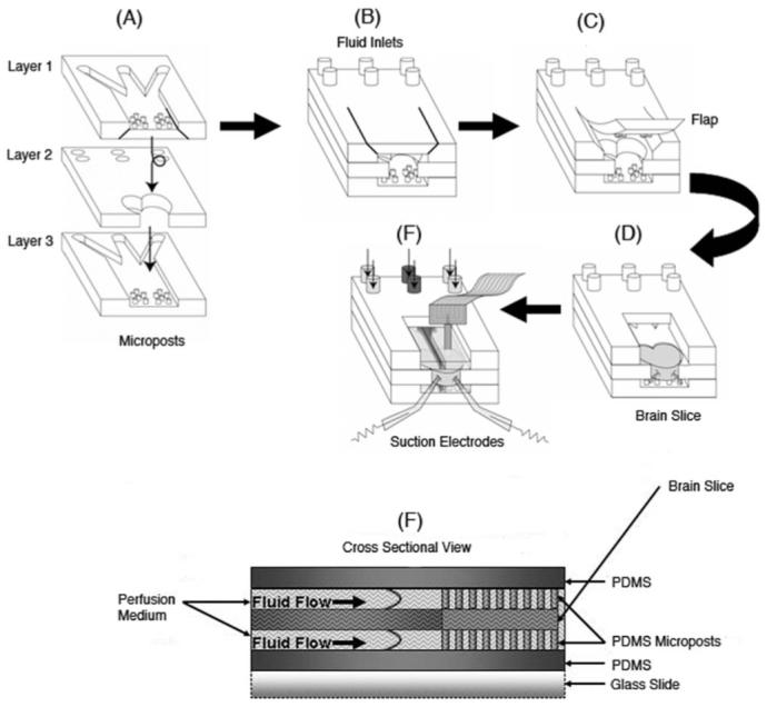
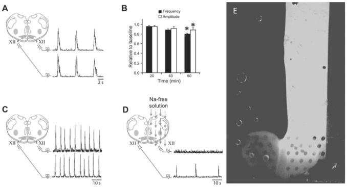
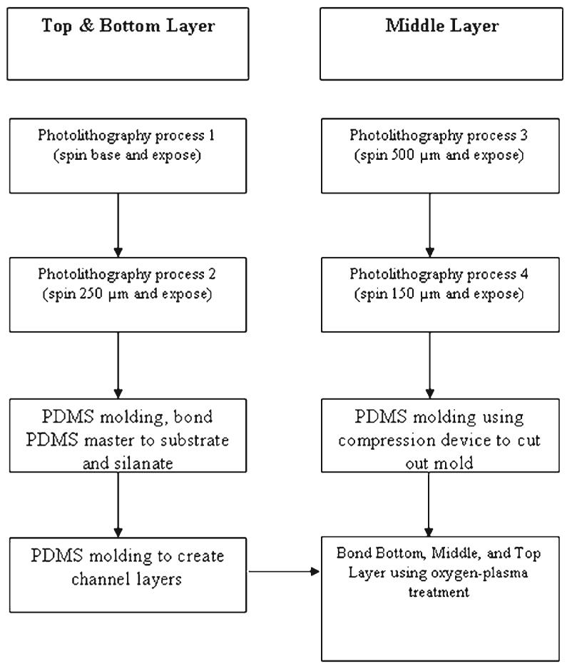
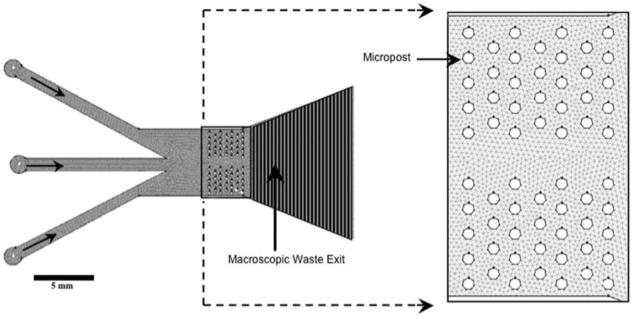
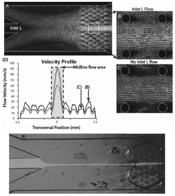
A novel three-layer microfluidic polydimethylsiloxane (PDMS) device was constructed with two fluid chambers that holds a brain slice in place with microposts while maintaining laminar perfusate flow above and below the slice. Our fabrication technique permits rapid production of PDMS layers that can be applied to brain slices of different shapes and sizes. In this study, the device was designed to fit the shape and thickness (530-700 microm) of a medullary brain slice taken from P0-P4 neonatal rats. Medullary slices in this chamber spontaneously produced rhythmic, respiratory-related motor output for up to 3 h, thereby demonstrating that brain slice viability was maintained for prolonged periods. This design is unique in that it achieves independent control of fluids through multiple channels in two separate fluid chambers. The laminar flow exhibited by the microfluidic chamber allows controlled solutions to target specific areas of the brain slice based on the input flow rates. To demonstrate this capability, a stream of Na(+)-free solution was focused on one half of a medullary slice to abolish spontaneous neural activity in only that half of the brain slice, while the other half remained active. We also demonstrated that flow of different solutions can be focused over the midline of the brain slice. The multilayer brain slice chamber design can integrate several traditional types of electrophysiology tools that are commonly used to measure neurophysiological properties of brain slices. Thus, this new microfluidic chamber is advantageous for experiments that involve controlled drug or solution delivery at high spatiotemporal resolution.
构建了一种新型的三层微流控聚二甲基硅氧烷(PDMS)装置,该装置有两个流体腔室,通过微柱将脑片固定在适当位置,同时在脑片上方和下方保持层流灌注液流动。我们的制造技术允许快速生产可应用于不同形状和大小脑片的PDMS层。在本研究中,该装置设计为适合取自出生后0至4天新生大鼠的延髓脑片的形状和厚度(530 - 700微米)。该腔室内的延髓脑片可自发产生有节律的、与呼吸相关的运动输出,持续长达3小时,从而证明脑片活力可长时间维持。这种设计的独特之处在于它通过两个独立流体腔室中的多个通道实现对流体的独立控制。微流控腔室呈现的层流允许基于输入流速将受控溶液靶向脑片的特定区域。为证明这一能力,将无钠溶液流聚焦在延髓脑片的一半上,仅使该半片脑片的自发神经活动消失,而另一半仍保持活跃。我们还证明了不同溶液流可聚焦在脑片的中线之上。多层脑片腔室设计可整合几种常用于测量脑片神经生理特性的传统类型的电生理工具。因此,这种新型微流控腔室对于涉及以高时空分辨率进行受控药物或溶液递送的实验具有优势。