Siebers M C, Walboomers X F, van den Dolder J, Leeuwenburgh S C G, Wolke J G C, Jansen J A
Department of Periodontology and Biomaterials, College of Dental Science 309, Radboud University Nijmegen Medical Centre, Nijmegen, The Netherlands.
J Mater Sci Mater Med. 2008 Feb;19(2):861-8. doi: 10.1007/s10856-007-0166-6. Epub 2007 Jul 31.
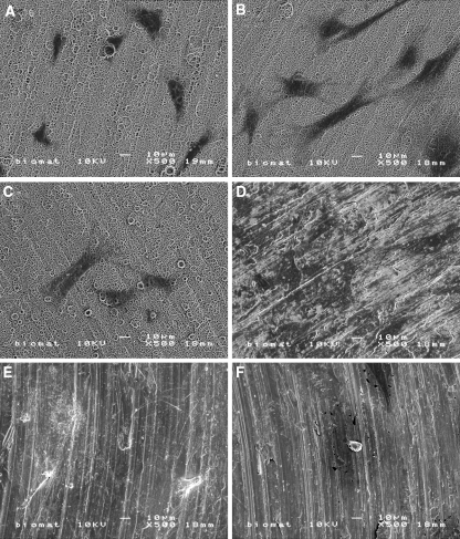
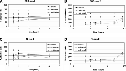
This study was designed to examine the influence of integrin subunit-beta1 and subunit-beta3 on the behavior of primary osteoblast-like cells, cultured on calcium phosphate (CaP)-coated and non coated titanium (Ti). Osteoblast-like cells were incubated with specific monoclonal antibodies against integrin-beta1 and integrin-beta3 to block the integrin function. Subsequently, cells were seeded on Ti discs, either non coated or provided with a 2 microm carbonated hydroxyapatite coating using Electrostatic Spray Deposition. Results showed that on CaP coatings, cellular attachment was decreased after a pre-treatment with either anti-integrin-beta1 or anti-integrin-beta3 antibodies. On Ti, cell adhesion was only slightly affected after a pre-treatment with anti-integrin-beta3 antibodies. Scanning electron microscopy showed that on both types of substrate, cellular morphology was not changed after a pre-treatment with either antibody. With quantitative PCR, it was shown for both substrates that mRNA expression of integrin-beta1 was increased after a pre-treatment with either anti-integrin-beta1 or anti-integrin-beta3 antibodies. Furthermore, after a pre-treatment with either antibody, mRNA expression of integrin-beta3 and ALP was decreased, on both types of substrate. In conclusion, osteoblast-like cells have the ability to compensate to great extent for the blocking strategy as applied here. Still, integrin-beta1 and beta3 seem to play different roles in attachment, proliferation, and differentiation of osteoblast-like cells, and responses on CaP-coated substrates differ to non coated Ti. Furthermore, the influence on ALP expression suggests involvement of both integrin subunits in signal transduction for cellular differentiation.
本研究旨在考察整合素β1亚基和β3亚基对原代成骨样细胞在磷酸钙(CaP)涂层和未涂层钛(Ti)上培养行为的影响。将成骨样细胞与抗整合素β1和抗整合素β3的特异性单克隆抗体孵育,以阻断整合素功能。随后,将细胞接种在未涂层的Ti盘或使用静电喷涂沉积提供2微米碳酸羟基磷灰石涂层的Ti盘上。结果表明,在CaP涂层上,用抗整合素β1或抗整合素β3抗体预处理后,细胞附着减少。在Ti上,用抗整合素β3抗体预处理后,细胞黏附仅受到轻微影响。扫描电子显微镜显示,在两种类型的底物上,用任何一种抗体预处理后,细胞形态均未改变。通过定量PCR表明,在两种底物上,用抗整合素β1或抗整合素β3抗体预处理后,整合素β1的mRNA表达均增加。此外,在两种底物上,用任何一种抗体预处理后,整合素β3和碱性磷酸酶(ALP)的mRNA表达均降低。总之,成骨样细胞有能力在很大程度上补偿此处应用的阻断策略。尽管如此,整合素β1和β3似乎在成骨样细胞的附着、增殖和分化中发挥不同作用,并且对CaP涂层底物的反应与未涂层的Ti不同。此外,对ALP表达的影响表明两种整合素亚基均参与细胞分化的信号转导。