Mukherjee Pratik, Hess Christopher P, Xu Duan, Han Eric T, Kelley Douglas A, Vigneron Daniel B
Department of Radiology, University of California-San Francisco, San Francisco, CA 94143-0628, USA.
Magn Reson Imaging. 2008 Feb;26(2):171-80. doi: 10.1016/j.mri.2007.05.011. Epub 2007 Aug 9.
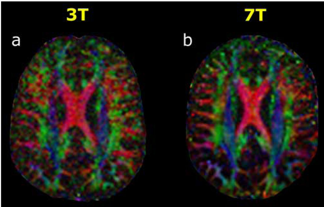
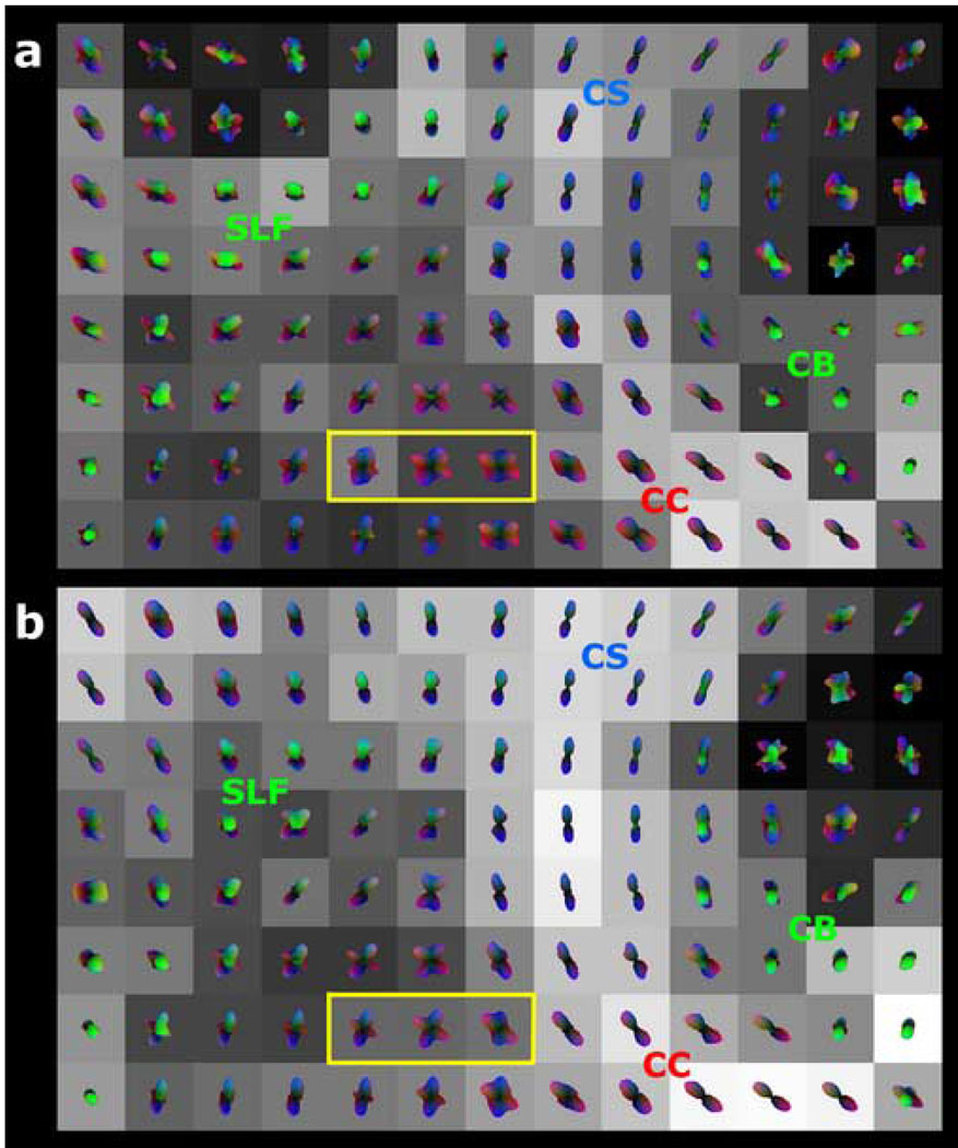
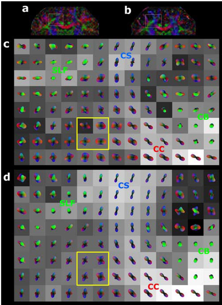
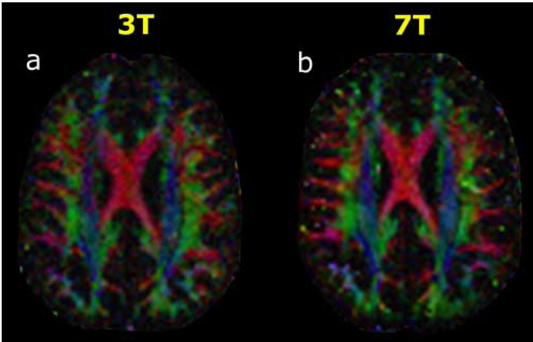
Diffusion tensor imaging (DTI) noninvasively depicts white matter connectivity in regions where the Gaussian model of diffusion is valid but yields inaccurate results in those where diffusion has a more complex distribution, such as fiber crossings. q-ball imaging (QBI) overcomes this limitation of DTI by more fully characterizing the angular dependence of intravoxel diffusion with larger numbers of diffusion-encoding directional measurements at higher diffusion-weighting factors (b values). However, the former technique results in longer acquisition times and the latter technique results in a lower signal-to-noise ratio (SNR). In this project, we developed specialized 7-T acquisition methods utilizing novel radiofrequency pulses, eight-channel parallel imaging EPI and high-order shimming with a phase-sensitive multichannel B0 field map reconstruction. These methods were applied in initial healthy adult volunteer studies, which demonstrated the feasibility of performing 7-T QBI. Preliminary comparisons of 3 T with 7 T within supratentorial crossing white matter tracts documented a 79.5% SNR increase for b=3000 s/mm2 (P=.0001) and a 38.6% SNR increase for b=6000 s/mm2 (P=.015). With spherical harmonic reconstruction of the q-ball orientation distribution function at b=3000 s/mm2, 7-T QBI allowed for accurate visualization of crossing fiber tracts with fewer diffusion-encoding acquisitions as compared with 3-T QBI. The improvement of 7-T QBI at b factors as high as 6000 s/mm2 resulted in better angular resolution as compared with 3-T QBI for depicting fibers crossing at shallow angles. Although the increased susceptibility effects at 7 T caused problematic distortions near brain-air interfaces at the skull base and posterior fossa, these initial 7-T QBI studies demonstrated excellent quality in much of the supratentorial brain, with significant improvements as compared with 3-T acquisitions in the same individuals.
扩散张量成像(DTI)可无创地描绘高斯扩散模型有效的区域中的白质连通性,但在扩散分布更为复杂的区域(如纤维交叉处)会产生不准确的结果。q球成像(QBI)通过在更高的扩散加权因子(b值)下进行更多数量的扩散编码方向测量,更全面地表征体素内扩散的角度依赖性,从而克服了DTI的这一局限性。然而,前一种技术会导致采集时间更长,而后一种技术会导致信噪比(SNR)更低。在本项目中,我们开发了专门的7T采集方法,利用新型射频脉冲、八通道并行成像EPI以及具有相敏多通道B0场图重建的高阶匀场技术。这些方法应用于最初的健康成人志愿者研究,证明了进行7T QBI的可行性。幕上交叉白质束内3T与7T的初步比较表明,对于b = 3000 s/mm2,SNR提高了79.5%(P = 0.0001),对于b = 6000 s/mm2,SNR提高了38.6%(P = 0.015)。在b = 3000 s/mm2时,通过q球方向分布函数的球谐重建,与3T QBI相比,7T QBI能够以更少的扩散编码采集准确显示交叉纤维束。与3T QBI相比,7T QBI在高达6000 s/mm2的b因子下的改进导致在描绘浅角度交叉纤维时具有更好的角度分辨率。尽管7T时增加的磁化率效应在颅底和后颅窝的脑气界面附近引起了有问题的畸变,但这些最初的7T QBI研究在幕上脑的大部分区域显示出了优异的质量,与同一受试者的3T采集相比有显著改善。