Smith Lester L, Niziolek Paul J, Haberstroh Karen M, Nauman Eric A, Webster Thomas J
Weldon School of Biomedical Engineering, Purdue University, West Lafayette, Indianapolis, IN, USA.
Int J Nanomedicine. 2007;2(3):383-8.
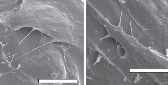
To facilitate locomotion and support the body, the skeleton relies on the transmission of forces between muscles and bones through complex junctions called entheses. The varying mechanical and biological properties of the enthesis make healing this avascular tissue difficult; hence the need for an engineered alternative. Cells in situ interact with their environment on the nano-scale which suggests that engineered approaches to enthesis regeneration should include such biologically-inspired nano-scale surface features. The present in vitro study investigated the effects of etching poly-lactic-co-glycolic acid (PLGA) scaffolds to produce nano-topography on the adhesion of fibroblasts and osteoblasts, two integral enthesis cell types. Nano-topography was produced on PLGA by etching the scaffolds in NaOH. Results showed that etching PLGA with NaOH to create nano-scale surface features decreased fibroblast adhesion while it increased osteoblast adhesion; criteria critical for the spatial control of osteoblast and fibroblast adhesion for a successful enthesis tissue engineering material. Thus, the results of this study showed for the first time collective evidence that PLGA can be either treated with NaOH or not on ends of an enthesis tissue engineering construct to spatially increase osteoblast and fibroblast adhesion, respectively.
为了便于身体移动和支撑身体,骨骼依靠肌肉和骨骼之间通过称为附着点的复杂连接来传递力量。附着点不同的机械和生物学特性使得修复这种无血管组织变得困难;因此需要一种工程替代方案。原位细胞在纳米尺度上与其环境相互作用,这表明附着点再生的工程方法应包括这种受生物启发的纳米尺度表面特征。目前的体外研究调查了蚀刻聚乳酸-乙醇酸共聚物(PLGA)支架以产生纳米拓扑结构对成纤维细胞和成骨细胞(两种重要的附着点细胞类型)黏附的影响。通过在NaOH中蚀刻支架在PLGA上产生纳米拓扑结构。结果表明,用NaOH蚀刻PLGA以创建纳米尺度表面特征会降低成纤维细胞的黏附,而增加成骨细胞的黏附;这对于成功的附着点组织工程材料中成骨细胞和成纤维细胞黏附的空间控制至关重要。因此,本研究结果首次共同证明,在附着点组织工程构建体的末端,PLGA可以分别通过用或不用NaOH处理来在空间上增加成骨细胞和成纤维细胞的黏附。