Grünwald David, Martin Robert M, Buschmann Volker, Bazett-Jones David P, Leonhardt Heinrich, Kubitscheck Ulrich, Cardoso M Cristina
Institute of Physical and Theoretical Chemistry, Rheinische Friedrich-Wilhelms-University, 53115 Bonn, Germany.
Biophys J. 2008 Apr 1;94(7):2847-58. doi: 10.1529/biophysj.107.115014. Epub 2007 Dec 7.
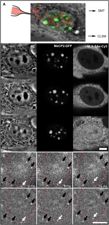
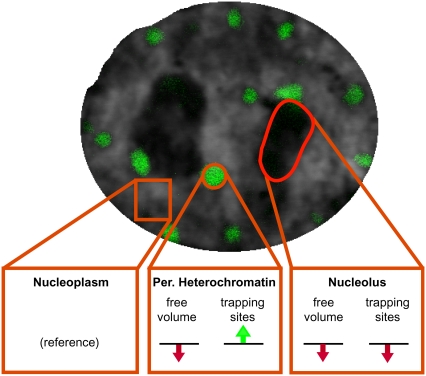
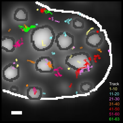
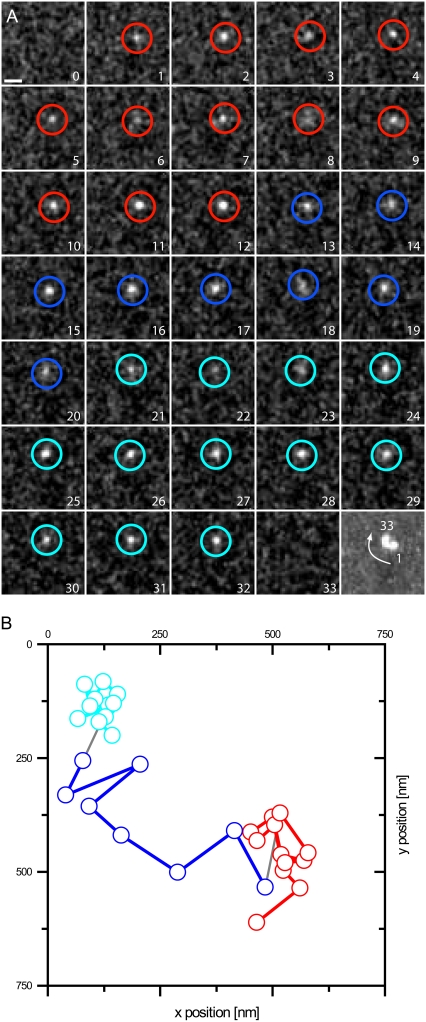
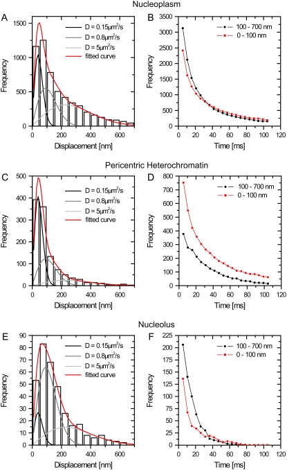
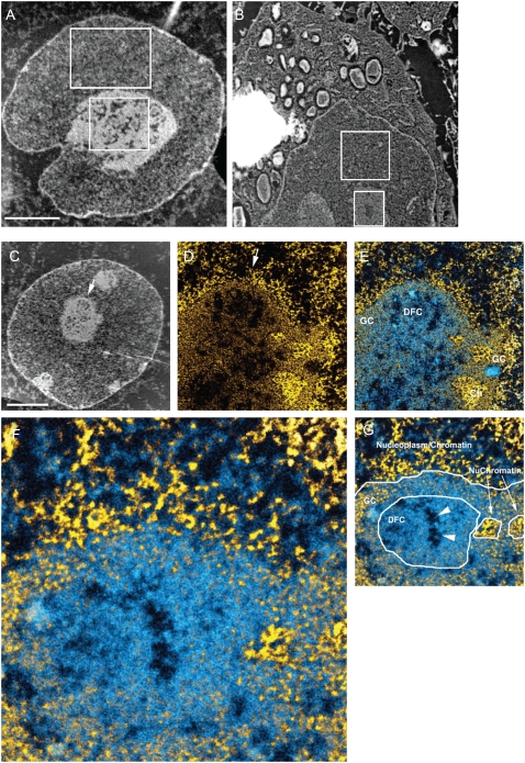
Genome activity and nuclear metabolism clearly depend on accessibility, but it is not known whether and to what extent nuclear structures limit the mobility and access of individual molecules. We used fluorescently labeled streptavidin with a nuclear localization signal as an average-sized, inert protein to probe the nuclear environment. The protein was injected into the cytoplasm of mouse cells, and single molecules were tracked in the nucleus with high-speed fluorescence microscopy. We analyzed and compared the mobility of single streptavidin molecules in structurally and functionally distinct nuclear compartments of living cells. Our results indicated that all nuclear subcompartments were easily and similarly accessible for such an average-sized protein, and even condensed heterochromatin neither excluded single molecules nor impeded their passage. The only significant difference was a higher frequency of transient trappings in heterochromatin, which lasted only tens of milliseconds. The streptavidin molecules, however, did not accumulate in heterochromatin, suggesting comparatively less free volume. Interestingly, the nucleolus seemed to exclude streptavidin, as it did many other nuclear proteins, when visualized by conventional fluorescence microscopy. The tracking of single molecules, nonetheless, showed no evidence for repulsion at the border but relatively unimpeded passage through the nucleolus. These results clearly show that single-molecule tracking can provide novel insights into mobility of proteins in the nucleus that cannot be obtained by conventional fluorescence microscopy. Our results suggest that nuclear processes may not be regulated at the level of physical accessibility but rather by local concentration of reactants and availability of binding sites.
基因组活性和核代谢显然依赖于可及性,但尚不清楚核结构是否以及在何种程度上限制了单个分子的移动性和可及性。我们使用带有核定位信号的荧光标记链霉亲和素作为一种中等大小的惰性蛋白来探测核环境。将该蛋白注射到小鼠细胞的细胞质中,并用高速荧光显微镜在细胞核中追踪单个分子。我们分析并比较了活细胞中结构和功能不同的核区室中单个链霉亲和素分子的移动性。我们的结果表明,对于这样一种中等大小的蛋白,所有核亚区室都易于且同样可及,甚至浓缩的异染色质既不排斥单个分子也不妨碍它们通过。唯一显著的差异是在异染色质中短暂捕获的频率较高,持续时间仅为几十毫秒。然而,链霉亲和素分子并未在异染色质中积累,这表明其自由体积相对较小。有趣的是,当通过传统荧光显微镜观察时,核仁似乎像排斥许多其他核蛋白一样排斥链霉亲和素。然而,单个分子的追踪并未显示在边界处有排斥的证据,而是表明其通过核仁的过程相对不受阻碍。这些结果清楚地表明单分子追踪能够为核内蛋白质的移动性提供传统荧光显微镜无法获得的新见解。我们的结果表明,核过程可能不是在物理可及性水平上受到调控,而是由反应物的局部浓度和结合位点的可用性来调控。