Shaik Zabeena P, Fifer E Kim, Nowak Grazyna
Department of Pharmaceutical Sciences, College of Pharmacy, University of Arkansas for Medical Sciences, Little Rock, Arkansas 72205, USA.
Am J Physiol Renal Physiol. 2008 Feb;294(2):F423-32. doi: 10.1152/ajprenal.00463.2007. Epub 2007 Dec 12.
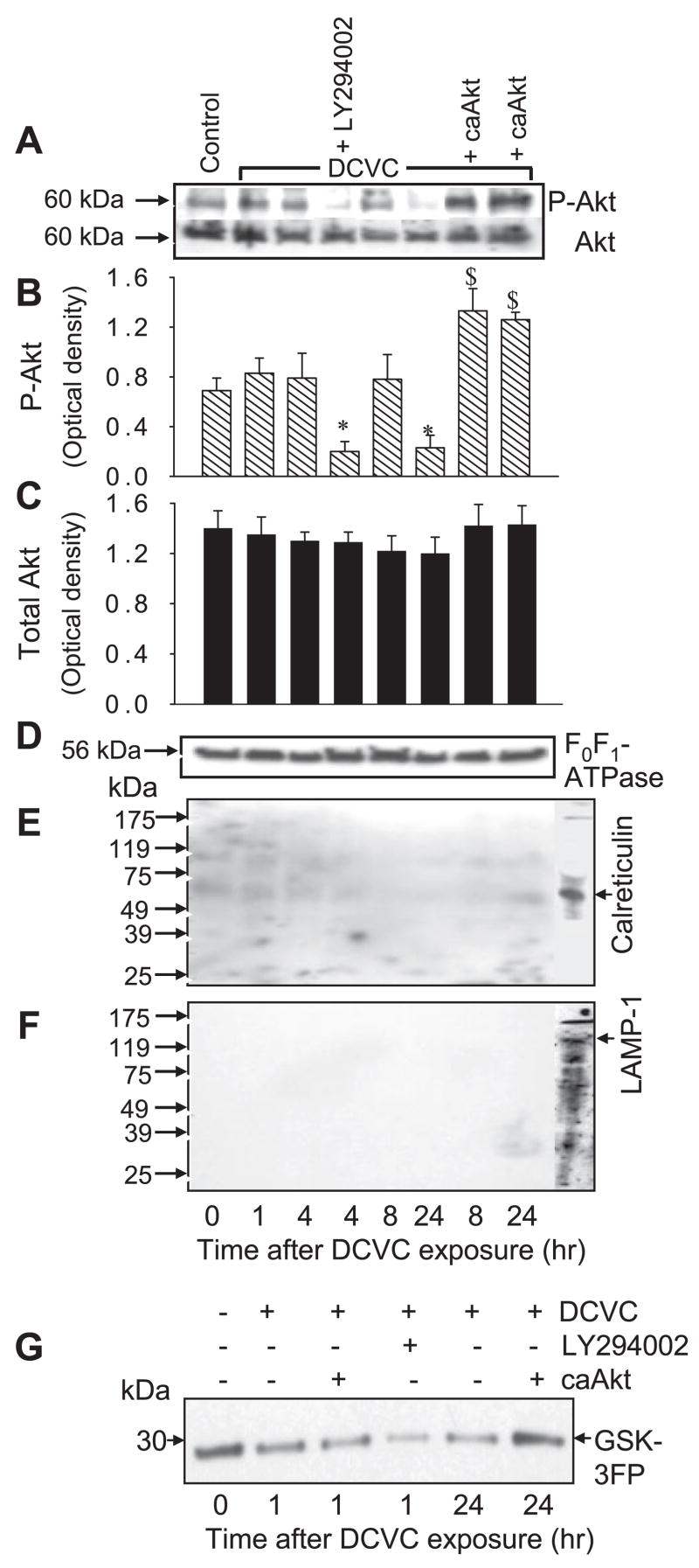
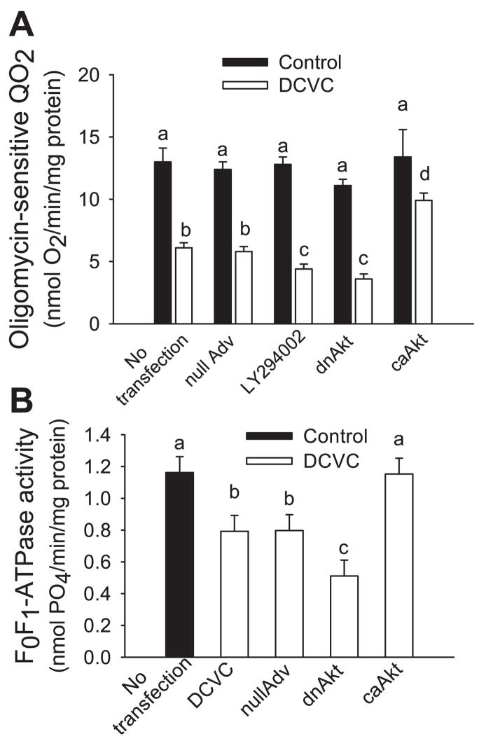
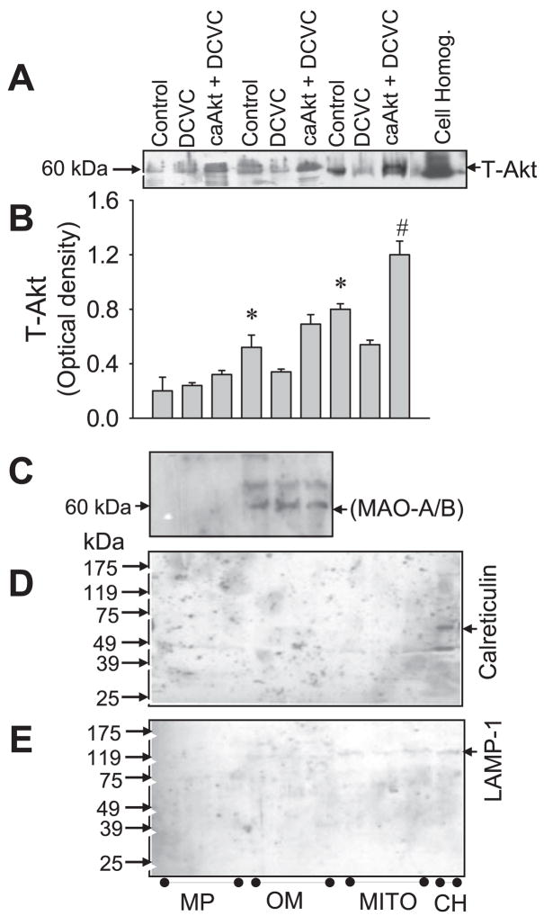
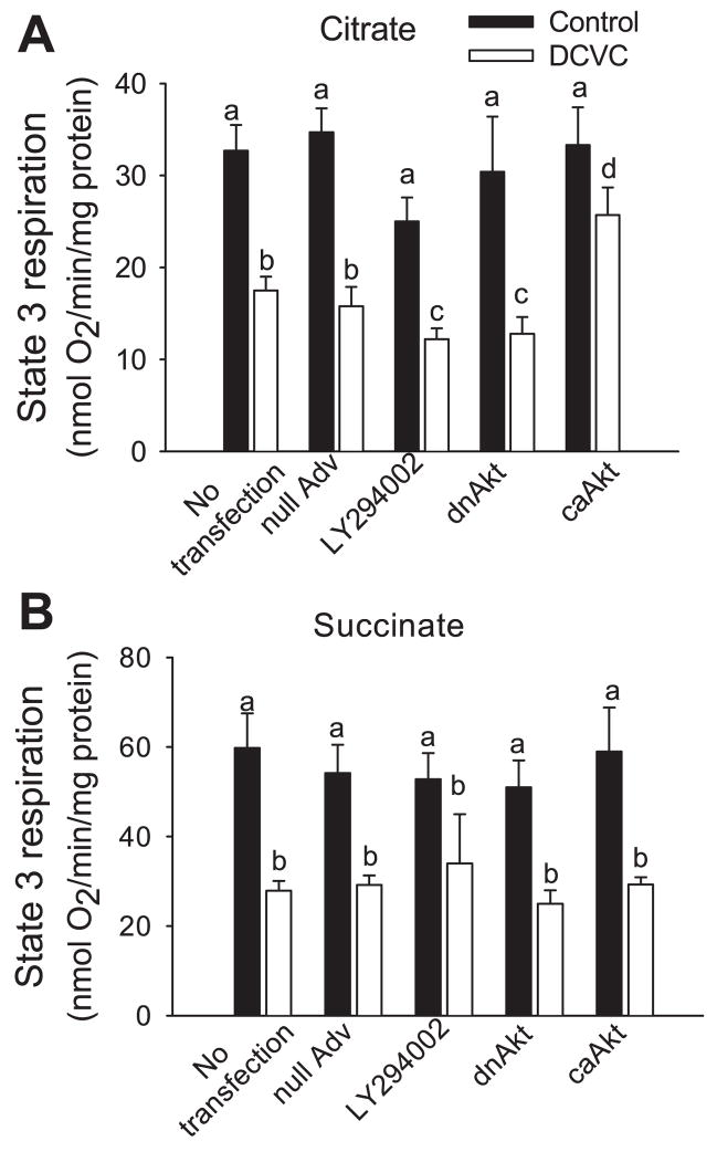
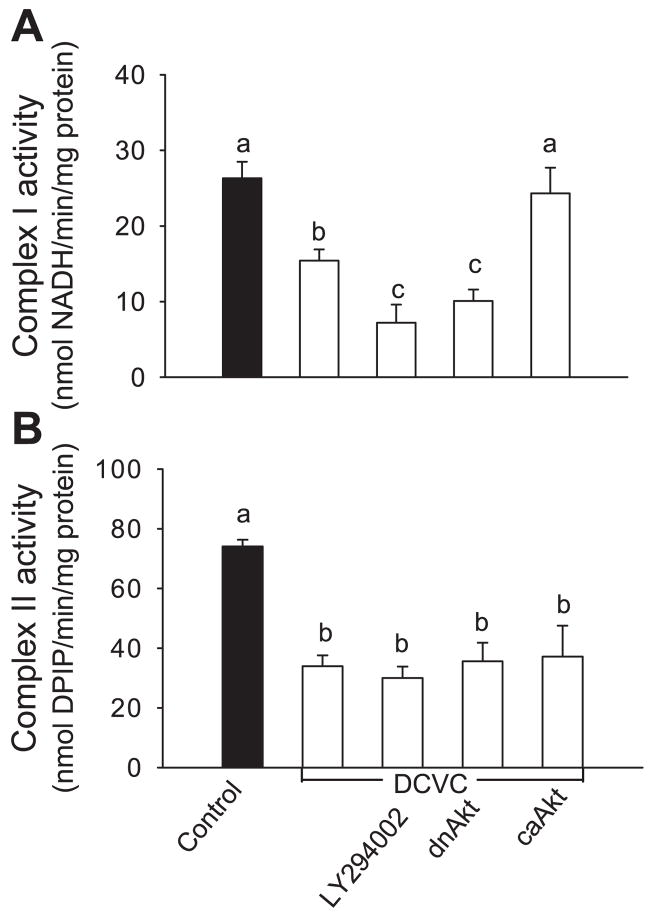
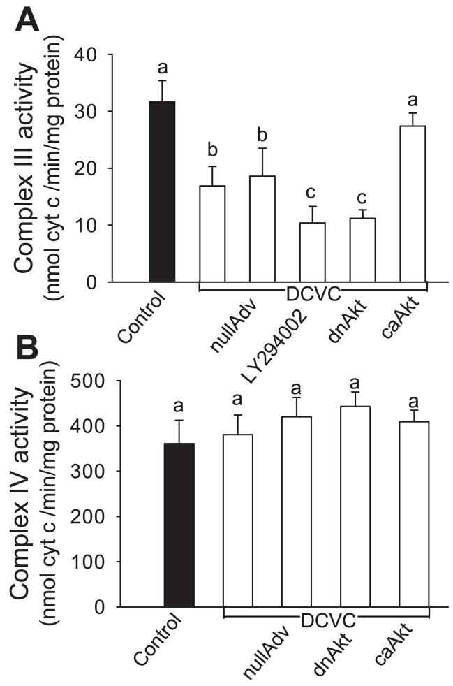
Previously, we showed that protein kinase B (Akt) activation increases intracellular ATP levels and decreases necrosis in renal proximal tubular cells (RPTC) injured by the nephrotoxicant S-(1, 2-dichlorovinyl)-l-cysteine (DCVC) (Shaik ZP, Fifer EK, Nowak G. Am J Physiol Renal Physiol 292: F292-F303, 2007). This study examined the role of Akt in improving mitochondrial function in DCVC-injured RPTC. Our data show a novel observation that phosphorylated (active) Akt is localized in mitochondria of noninjured RPTC, both in mitoplasts and the mitochondrial outer membrane. Mitochondrial levels of active Akt decreased in nephrotoxicant-injured RPTC, and this decrease was associated with mitochondrial dysfunction. DCVC decreased basal, uncoupled, and state 3 respirations; ATP production; activities of complexes I, II, and III; the mitochondrial membrane potential (DeltaPsi(m)); and F(0)F(1)-ATPase activity. Expressing constitutively active Akt in DCVC-injured RPTC increased the levels of phosphorylated Akt in mitochondria, reduced the decreases in basal and uncoupled respirations, increased complex I-coupled state 3 respiration and ATP production, enhanced activities of complex I, complex III, and F(0)F(1)-ATPase, and improved DeltaPsi(m). In contrast, inhibiting Akt activation by expressing dominant negative (inactive) Akt or using 20 microM LY294002 exacerbated decreases in electron transport rate, state 3 respiration, ATP production, DeltaPsi(m), and activities of complex I, complex III, and F(0)F(1)-ATPase. In conclusion, our data show that Akt activation promotes mitochondrial respiration and ATP production in toxicant-injured RPTC by 1) improving integrity of the respiratory chain and maintaining activities of complex I and complex III, 2) reducing decreases in DeltaPsi(m), and 3) restoring F(0)F(1)-ATPase activity.
此前,我们发现蛋白激酶B(Akt)激活可提高细胞内ATP水平,并减少由肾毒性物质S-(1,2-二氯乙烯基)-L-半胱氨酸(DCVC)损伤的肾近端小管细胞(RPTC)的坏死(Shaik ZP,Fifer EK,Nowak G.《美国生理学杂志:肾脏生理学》292:F292 - F303,2007)。本研究检测了Akt在改善DCVC损伤的RPTC线粒体功能中的作用。我们的数据显示了一个新的发现,即磷酸化(活性)Akt定位于未损伤RPTC的线粒体中,存在于线粒体膜间隙和线粒体外膜。在肾毒性物质损伤的RPTC中,活性Akt的线粒体水平降低,且这种降低与线粒体功能障碍相关。DCVC降低了基础呼吸、解偶联呼吸和状态3呼吸;ATP生成;复合物I、II和III的活性;线粒体膜电位(ΔΨm);以及F0F1 - ATP酶活性。在DCVC损伤的RPTC中表达组成型活性Akt可增加线粒体中磷酸化Akt的水平,减少基础呼吸和解偶联呼吸的降低,增加复合物I偶联的状态3呼吸和ATP生成,增强复合物I、复合物III和F0F1 - ATP酶的活性,并改善ΔΨm。相反,通过表达显性负性(无活性)Akt或使用20μM LY294002抑制Akt激活会加剧电子传递速率、状态3呼吸、ATP生成、ΔΨm以及复合物I、复合物III和F0F1 - ATP酶活性的降低。总之:我们的数据表明,Akt激活通过以下方式促进中毒损伤的RPTC中的线粒体呼吸和ATP生成:1)改善呼吸链的完整性并维持复合物I和复合物III的活性;2)减少ΔΨm的降低;3)恢复F0F1 - ATP酶活性。