• 文献检索
  • 文档翻译
  • 深度研究
  • 学术资讯
  • Suppr Zotero 插件Zotero 插件
  • 邀请有礼
  • 套餐&价格
  • 历史记录
应用&插件
Suppr Zotero 插件Zotero 插件浏览器插件Mac 客户端Windows 客户端微信小程序
定价
高级版会员购买积分包购买API积分包
服务
文献检索文档翻译深度研究API 文档MCP 服务
关于我们
关于 Suppr公司介绍联系我们用户协议隐私条款
关注我们

Suppr 超能文献

核心技术专利:CN118964589B侵权必究
粤ICP备2023148730 号-1Suppr @ 2026

文献检索

告别复杂PubMed语法,用中文像聊天一样搜索,搜遍4000万医学文献。AI智能推荐,让科研检索更轻松。

立即免费搜索

文件翻译

保留排版,准确专业,支持PDF/Word/PPT等文件格式,支持 12+语言互译。

免费翻译文档

深度研究

AI帮你快速写综述,25分钟生成高质量综述,智能提取关键信息,辅助科研写作。

立即免费体验

相似文献

1
Restricted lateral mobility of plasma membrane CD4 impairs HIV-1 envelope glycoprotein mediated fusion.质膜CD4的侧向移动受限会损害HIV-1包膜糖蛋白介导的融合。
Mol Membr Biol. 2008 Jan;25(1):83-94. doi: 10.1080/09687680701613713.
2
The role of cholesterol and sphingolipids in chemokine receptor function and HIV-1 envelope glycoprotein-mediated fusion.胆固醇和鞘脂在趋化因子受体功能及HIV-1包膜糖蛋白介导的融合中的作用
Virol J. 2006 Dec 22;3:104. doi: 10.1186/1743-422X-3-104.
3
Functional expression of CD4, CXCR4, and CCR5 in glycosphingolipid-deficient mouse melanoma GM95 cells and susceptibility to HIV-1 envelope glycoprotein-triggered membrane fusion.CD4、CXCR4和CCR5在鞘糖脂缺陷型小鼠黑色素瘤GM95细胞中的功能表达及对HIV-1包膜糖蛋白触发的膜融合的敏感性。
Virology. 2004 Jan 5;318(1):55-65. doi: 10.1016/j.virol.2003.08.042.
4
Coreceptor competition for association with CD4 may change the susceptibility of human cells to infection with T-tropic and macrophagetropic isolates of human immunodeficiency virus type 1.与CD4结合的共受体竞争可能会改变人类细胞对1型人类免疫缺陷病毒的T嗜性和巨噬细胞嗜性分离株感染的易感性。
J Virol. 2000 Jun;74(11):5016-23. doi: 10.1128/jvi.74.11.5016-5023.2000.
5
Fusion of HIV-1 envelope-expressing cells to human glomerular endothelial cells through an CXCR4-mediated mechanism.通过CXCR4介导的机制,HIV-1包膜表达细胞与人类肾小球内皮细胞融合。
Pediatr Nephrol. 2005 Oct;20(10):1401-9. doi: 10.1007/s00467-005-1950-5. Epub 2005 Jul 27.
6
Conserved changes in envelope function during human immunodeficiency virus type 1 coreceptor switching.人类免疫缺陷病毒1型共受体转换过程中包膜功能的保守变化。
J Virol. 2007 Aug;81(15):8165-79. doi: 10.1128/JVI.02792-06. Epub 2007 May 16.
7
Promonocytic U937 subclones expressing CD4 and CXCR4 are resistant to infection with and cell-to-cell fusion by T-cell-tropic human immunodeficiency virus type 1.表达CD4和CXCR4的单核细胞U937亚克隆对嗜T细胞性1型人类免疫缺陷病毒的感染及细胞间融合具有抗性。
J Virol. 1997 Dec;71(12):9664-71. doi: 10.1128/JVI.71.12.9664-9671.1997.
8
Engineered CD4- and CXCR4-using simian immunodeficiency virus from African green monkeys is neutralization sensitive and replicates in nonstimulated lymphocytes.利用来自非洲绿猴的猿猴免疫缺陷病毒构建的CD4和CXCR4对中和敏感,且能在未受刺激的淋巴细胞中复制。
J Virol. 2002 Nov;76(21):10627-36. doi: 10.1128/jvi.76.21.10627-10636.2002.
9
Activation and Inactivation of Primary Human Immunodeficiency Virus Envelope Glycoprotein Trimers by CD4-Mimetic Compounds.CD4模拟化合物对原代人免疫缺陷病毒包膜糖蛋白三聚体的激活与失活作用
J Virol. 2017 Jan 18;91(3). doi: 10.1128/JVI.01880-16. Print 2017 Feb 1.
10
Envelope glycoproteins from human immunodeficiency virus types 1 and 2 and simian immunodeficiency virus can use human CCR5 as a coreceptor for viral entry and make direct CD4-dependent interactions with this chemokine receptor.1型和2型人类免疫缺陷病毒以及猿猴免疫缺陷病毒的包膜糖蛋白可将人类CCR5用作病毒进入的共受体,并与这种趋化因子受体进行直接的CD4依赖性相互作用。
J Virol. 1997 Sep;71(9):6296-304. doi: 10.1128/JVI.71.9.6296-6304.1997.

引用本文的文献

1
Analysis of Factors That Regulate HIV-1 Fusion in Reverse.HIV-1逆转融合调控因素分析
Viruses. 2025 Mar 26;17(4):472. doi: 10.3390/v17040472.
2
HIV and SIV Envelope Glycoproteins Interact with Glycolipids and Lipids.HIV 和 SIV 包膜糖蛋白与糖脂和脂质相互作用。
Int J Mol Sci. 2023 Jul 21;24(14):11730. doi: 10.3390/ijms241411730.
3
The Manifold Roles of Sphingolipids in Viral Infections.鞘脂类在病毒感染中的多种作用
Front Physiol. 2021 Sep 29;12:715527. doi: 10.3389/fphys.2021.715527. eCollection 2021.
4
Sphingolipids: Effectors and Achilles Heals in Viral Infections?鞘脂类:病毒感染中的效应因子和阿喀琉斯之踵?
Cells. 2021 Aug 24;10(9):2175. doi: 10.3390/cells10092175.
5
A biophysical perspective on receptor-mediated virus entry with a focus on HIV.从生物物理角度看受体介导的病毒进入,重点是 HIV。
Biochim Biophys Acta Biomembr. 2020 Jun 1;1862(6):183158. doi: 10.1016/j.bbamem.2019.183158. Epub 2019 Dec 19.
6
Membrane Dynamics in Health and Disease: Impact on Cellular Signalling.膜动力学在健康和疾病中的作用:对细胞信号转导的影响。
J Membr Biol. 2019 Oct;252(4-5):213-226. doi: 10.1007/s00232-019-00087-0. Epub 2019 Aug 21.
7
Modeling bispecific monoclonal antibody interaction with two cell membrane targets indicates the importance of surface diffusion.对双特异性单克隆抗体与两个细胞膜靶点相互作用进行建模表明了表面扩散的重要性。
MAbs. 2016 Jul;8(5):905-15. doi: 10.1080/19420862.2016.1178437. Epub 2016 Apr 20.
8
Membrane organization of virus and target cell plays a role in HIV entry.病毒和靶细胞的膜组织在HIV进入过程中发挥作用。
Biochimie. 2014 Dec;107 Pt A:22-7. doi: 10.1016/j.biochi.2014.08.015. Epub 2014 Sep 1.
9
Amphotericin B increases influenza A virus infection by preventing IFITM3-mediated restriction.两性霉素 B 通过阻止 IFITM3 介导的限制来增加甲型流感病毒感染。
Cell Rep. 2013 Nov 27;5(4):895-908. doi: 10.1016/j.celrep.2013.10.033. Epub 2013 Nov 21.
10
Thermal stability of the human immunodeficiency virus type 1 (HIV-1) receptors, CD4 and CXCR4, reconstituted in proteoliposomes.人免疫缺陷病毒 1 型(HIV-1)受体 CD4 和 CXCR4 在脂蛋白体中重建的热稳定性。
PLoS One. 2010 Oct 13;5(10):e13249. doi: 10.1371/journal.pone.0013249.

本文引用的文献

1
Sphingomyelinase restricts the lateral diffusion of CD4 and inhibits human immunodeficiency virus fusion.鞘磷脂酶限制CD4的侧向扩散并抑制人类免疫缺陷病毒融合。
J Virol. 2007 May;81(10):5294-304. doi: 10.1128/JVI.02553-06. Epub 2007 Mar 7.
2
Effect of cholesterol on lateral diffusion of fluorescent lipid probes in native hippocampal membranes.胆固醇对天然海马体膜中荧光脂质探针横向扩散的影响。
Chem Phys Lipids. 2006 Sep;143(1-2):11-21. doi: 10.1016/j.chemphyslip.2006.04.003. Epub 2006 May 7.
3
Cellular entry of HIV: Evaluation of therapeutic targets.HIV的细胞进入:治疗靶点评估
Curr Pharm Des. 2006;12(16):1963-73. doi: 10.2174/138161206777442155.
4
Sphingolipids, cholesterol, and HIV-1: a paradigm in viral fusion.鞘脂、胆固醇与HIV-1:病毒融合的范例
Glycoconj J. 2006 May;23(3-4):189-97. doi: 10.1007/s10719-006-7924-4.
5
Sphingolipids: modulators of HIV-1 infection and pathogenesis.鞘脂类:HIV-1感染及发病机制的调节因子
Biosci Rep. 2005 Oct-Dec;25(5-6):329-43. doi: 10.1007/s10540-005-2894-5.
6
Regulation of human immunodeficiency virus type 1 envelope glycoprotein fusion by a membrane-interactive domain in the gp41 cytoplasmic tail.通过gp41胞质尾部的膜相互作用结构域对1型人类免疫缺陷病毒包膜糖蛋白融合的调控。
J Virol. 2005 Oct;79(19):12231-41. doi: 10.1128/JVI.79.19.12231-12241.2005.
7
Dynamic reorganization of chemokine receptors, cholesterol, lipid rafts, and adhesion molecules to sites of CD4 engagement.趋化因子受体、胆固醇、脂筏和黏附分子向CD4结合位点的动态重组。
Exp Cell Res. 2005 Apr 1;304(2):559-69. doi: 10.1016/j.yexcr.2004.11.022. Epub 2004 Dec 23.
8
Ceramide, a target for antiretroviral therapy.神经酰胺,一种抗逆转录病毒疗法的靶点。
Proc Natl Acad Sci U S A. 2004 Oct 26;101(43):15452-7. doi: 10.1073/pnas.0402874101. Epub 2004 Oct 15.
9
The role of lipid microdomains in virus biology.脂筏在病毒生物学中的作用。
Subcell Biochem. 2004;37:443-91. doi: 10.1007/978-1-4757-5806-1_14.
10
Mobility of the human immunodeficiency virus (HIV) receptor CD4 and coreceptor CCR5 in living cells: implications for HIV fusion and entry events.人类免疫缺陷病毒(HIV)受体CD4和共受体CCR5在活细胞中的流动性:对HIV融合和进入事件的影响。
J Virol. 2004 Sep;78(17):9573-8. doi: 10.1128/JVI.78.17.9573-9578.2004.

质膜CD4的侧向移动受限会损害HIV-1包膜糖蛋白介导的融合。

Restricted lateral mobility of plasma membrane CD4 impairs HIV-1 envelope glycoprotein mediated fusion.

作者信息

Rawat Satinder S, Zimmerman Christina, Johnson Benitra T, Cho Edward, Lockett Stephen J, Blumenthal Robert, Puri Anu

机构信息

CCRNP, NCI-Frederick, National Institutes of Health, Frederick, Maryland 21702-1201, USA.

出版信息

Mol Membr Biol. 2008 Jan;25(1):83-94. doi: 10.1080/09687680701613713.

DOI:10.1080/09687680701613713
PMID:18097956
原文链接:https://pmc.ncbi.nlm.nih.gov/articles/PMC3466082/
Abstract

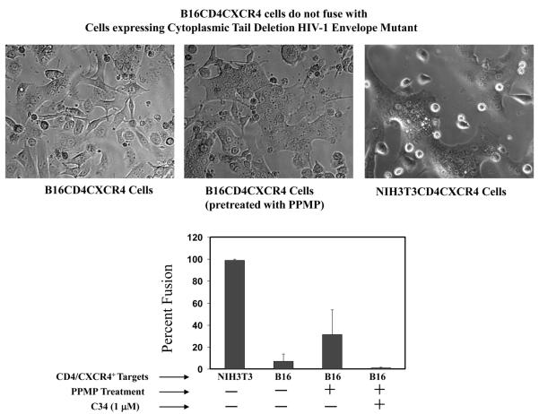
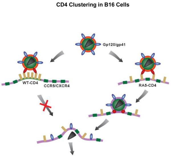
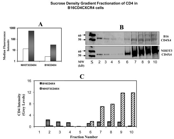
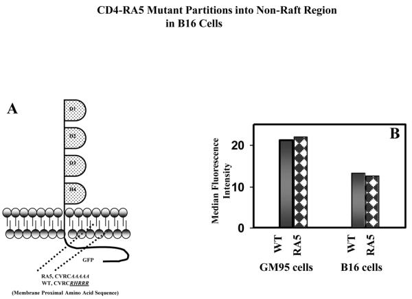
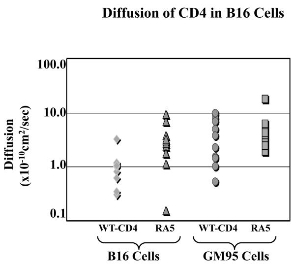
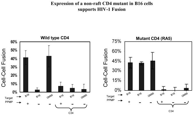
We investigated the effect of receptor mobility on HIV-1 envelope glycoprotein (Env)-triggered fusion using B16 mouse melanoma cells that are engineered to express CD4 and CXCR4 or CCR5. These engineered cells are resistant to fusion mediated CD4-dependent HIV-1 envelope glycoprotein. Receptor mobility was measured by fluorescence recovery after photobleaching (FRAP) using either fluorescently-labeled antibodies or transient expression of GFP-tagged receptors in the cells. No significant differences between B16 and NIH3T3 (fusion-permissive) cells were seen in lateral mobility of CCR5 or lipid probes. By contrast CD4 mobility in B16 cells was about seven-fold reduced compared to its mobility in fusion-permissive NIH3T3 cells. However, a CD4 mutant (RA5) that localizes to non-raft membrane microdomains exhibited a three-fold increased mobility in B16 cells as compared with WT-CD4. Interestingly, the B16 cells expressing the RA5 mutant (but not the wild type CD4) and coreceptors supported HIV-1 Env-mediated fusion. Our data demonstrate that the lateral mobility of CD4 is an important determinant of HIV-1 fusion/entry.

摘要

我们使用经过基因工程改造以表达CD4和CXCR4或CCR5的B16小鼠黑色素瘤细胞,研究了受体流动性对HIV-1包膜糖蛋白(Env)触发融合的影响。这些经过基因工程改造的细胞对融合介导的CD4依赖性HIV-1包膜糖蛋白具有抗性。通过使用荧光标记抗体或细胞中GFP标记受体的瞬时表达,通过光漂白后荧光恢复(FRAP)测量受体流动性。在CCR5或脂质探针的横向流动性方面,未观察到B16细胞与NIH3T3(融合许可)细胞之间存在显著差异。相比之下,B16细胞中CD4的流动性与其在融合许可的NIH3T3细胞中的流动性相比降低了约7倍。然而,定位于非脂筏膜微区的CD4突变体(RA5)在B16细胞中的流动性与野生型CD4相比增加了3倍。有趣的是,表达RA5突变体(而非野生型CD4)和共受体的B16细胞支持HIV-1 Env介导的融合。我们的数据表明,CD4的横向流动性是HIV-1融合/进入的重要决定因素。

https://cdn.ncbi.nlm.nih.gov/pmc/blobs/d3b0/3466082/a8a386627714/nihms-341310-f0007.jpg
https://cdn.ncbi.nlm.nih.gov/pmc/blobs/d3b0/3466082/f35f9f1a9dac/nihms-341310-f0001.jpg
https://cdn.ncbi.nlm.nih.gov/pmc/blobs/d3b0/3466082/d30a3f6774ce/nihms-341310-f0002.jpg
https://cdn.ncbi.nlm.nih.gov/pmc/blobs/d3b0/3466082/0dd4730b8810/nihms-341310-f0003.jpg
https://cdn.ncbi.nlm.nih.gov/pmc/blobs/d3b0/3466082/d2fb56756486/nihms-341310-f0005.jpg
https://cdn.ncbi.nlm.nih.gov/pmc/blobs/d3b0/3466082/022ac1303ba0/nihms-341310-f0006.jpg
https://cdn.ncbi.nlm.nih.gov/pmc/blobs/d3b0/3466082/a8a386627714/nihms-341310-f0007.jpg