• 文献检索
  • 文档翻译
  • 深度研究
  • 学术资讯
  • Suppr Zotero 插件Zotero 插件
  • 邀请有礼
  • 套餐&价格
  • 历史记录
应用&插件
Suppr Zotero 插件Zotero 插件浏览器插件Mac 客户端Windows 客户端微信小程序
定价
高级版会员购买积分包购买API积分包
服务
文献检索文档翻译深度研究API 文档MCP 服务
关于我们
关于 Suppr公司介绍联系我们用户协议隐私条款
关注我们

Suppr 超能文献

核心技术专利:CN118964589B侵权必究
粤ICP备2023148730 号-1Suppr @ 2026

文献检索

告别复杂PubMed语法,用中文像聊天一样搜索,搜遍4000万医学文献。AI智能推荐,让科研检索更轻松。

立即免费搜索

文件翻译

保留排版,准确专业,支持PDF/Word/PPT等文件格式,支持 12+语言互译。

免费翻译文档

深度研究

AI帮你快速写综述,25分钟生成高质量综述,智能提取关键信息,辅助科研写作。

立即免费体验

生物力学信号对软骨细胞基因表达的调控

Regulation of chondrocytic gene expression by biomechanical signals.

作者信息

Knobloch Thomas J, Madhavan Shashi, Nam Jin, Agarwal Suresh, Agarwal Sudha

机构信息

Biomechanics and Tissue Engineering Laboratory, Section of Oral Biology, Ohio State University College of Dentistry, 305 West 12th Avenue, Columbus, OH 43210, USA.

出版信息

Crit Rev Eukaryot Gene Expr. 2008;18(2):139-50. doi: 10.1615/critreveukargeneexpr.v18.i2.30.

DOI:10.1615/critreveukargeneexpr.v18.i2.30
PMID:18304028
原文链接:https://pmc.ncbi.nlm.nih.gov/articles/PMC4967411/
Abstract

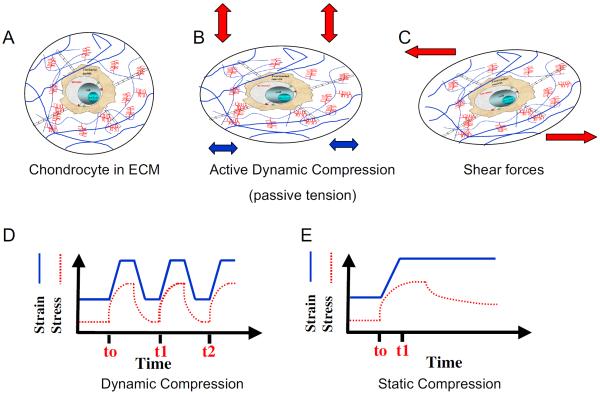
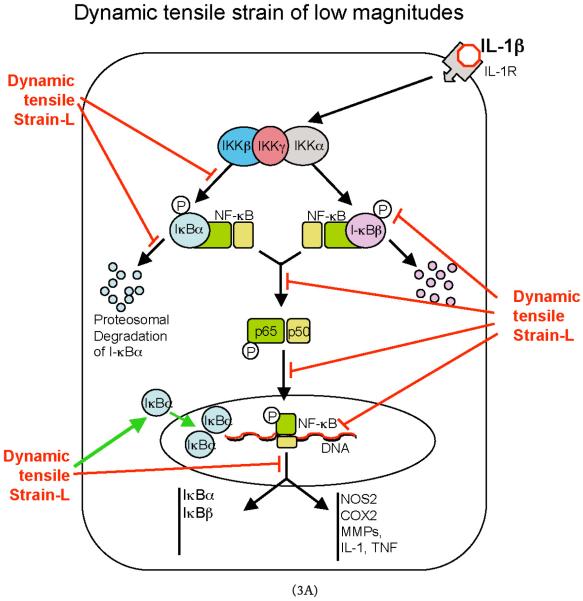
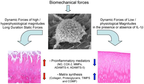
Cartilage is a mechanosensitive tissue, which means that it can perceive and respond to biomechanical signals. Despite the known importance of biomechanical signals in the etiopathogenesis of arthritic diseases and their effectiveness in joint restoration, little is understood about their actions at the cellular level. Recent molecular approaches have revealed that specific biomechanical stimuli and cell interactions generate intracellular signals that are powerful inducers or suppressors of proinflammatory and reparative genes in chondrocytes. Biomechanical signals are perceived by cartilage in magnitude-, frequency-, and time-dependent manners. Static and dynamic biomechanical forces of high magnitudes induce proinflammatory genes and inhibit matrix synthesis. Contrarily, dynamic biomechanical signals of low/physiologic magnitudes are potent antiinflammatory signals that inhibit interleukin-1beta (IL-1beta)-induced proinflammatory gene transcription and abrogate IL-1beta/tumor necrosis factor-alpha-induced inhibition of matrix synthesis. Recent studies have identified nuclear factor-kB (NF-kB) transcription factors as key regulators of biomechanical signal-mediated proinflammatory and antiinflammatory actions. These signals intercept multiple steps in the NF-kappaB signaling cascade to regulate cytokine gene expression. Taken together, these findings provide insight into how biomechanical signals regulate inflammatory and reparative gene transcription, underscoring their potential in enhancing the ability of chondrocytes to curb inflammation in diseased joints.

摘要

软骨是一种机械敏感组织,这意味着它能够感知并对生物力学信号做出反应。尽管生物力学信号在关节炎性疾病的发病机制中具有已知的重要性,并且在关节修复中也有效果,但对于它们在细胞水平上的作用却了解甚少。最近的分子方法揭示,特定的生物力学刺激和细胞相互作用会产生细胞内信号,这些信号是软骨细胞中促炎和修复基因的强大诱导剂或抑制剂。软骨以大小、频率和时间依赖性方式感知生物力学信号。高强度的静态和动态生物力学力会诱导促炎基因并抑制基质合成。相反,低/生理强度的动态生物力学信号是有效的抗炎信号,可抑制白细胞介素-1β(IL-1β)诱导的促炎基因转录,并消除IL-1β/肿瘤坏死因子-α诱导的基质合成抑制。最近的研究已确定核因子-κB(NF-κB)转录因子是生物力学信号介导的促炎和抗炎作用的关键调节因子。这些信号在NF-κB信号级联反应的多个步骤中起作用,以调节细胞因子基因表达。综上所述,这些发现为生物力学信号如何调节炎症和修复基因转录提供了见解,强调了它们在增强软骨细胞抑制患病关节炎症能力方面的潜力。

https://cdn.ncbi.nlm.nih.gov/pmc/blobs/73de/4967411/02ef74d8da08/nihms-802028-f0003.jpg
https://cdn.ncbi.nlm.nih.gov/pmc/blobs/73de/4967411/edc9bef2a20f/nihms-802028-f0001.jpg
https://cdn.ncbi.nlm.nih.gov/pmc/blobs/73de/4967411/5e318ba059e0/nihms-802028-f0002.jpg
https://cdn.ncbi.nlm.nih.gov/pmc/blobs/73de/4967411/02ef74d8da08/nihms-802028-f0003.jpg
https://cdn.ncbi.nlm.nih.gov/pmc/blobs/73de/4967411/edc9bef2a20f/nihms-802028-f0001.jpg
https://cdn.ncbi.nlm.nih.gov/pmc/blobs/73de/4967411/5e318ba059e0/nihms-802028-f0002.jpg
https://cdn.ncbi.nlm.nih.gov/pmc/blobs/73de/4967411/02ef74d8da08/nihms-802028-f0003.jpg

相似文献

1
Regulation of chondrocytic gene expression by biomechanical signals.生物力学信号对软骨细胞基因表达的调控
Crit Rev Eukaryot Gene Expr. 2008;18(2):139-50. doi: 10.1615/critreveukargeneexpr.v18.i2.30.
2
Biomechanical thresholds regulate inflammation through the NF-kappaB pathway: experiments and modeling.生物力学阈值通过核因子κB通路调节炎症:实验与建模
PLoS One. 2009;4(4):e5262. doi: 10.1371/journal.pone.0005262. Epub 2009 Apr 16.
3
Biomechanical signals inhibit IKK activity to attenuate NF-kappaB transcription activity in inflamed chondrocytes.生物力学信号抑制IKK活性,以减弱炎症软骨细胞中的NF-κB转录活性。
Arthritis Rheum. 2007 Oct;56(10):3284-96. doi: 10.1002/art.22933.
4
Role of NF-kappaB transcription factors in antiinflammatory and proinflammatory actions of mechanical signals.核因子-κB转录因子在机械信号的抗炎和促炎作用中的作用
Arthritis Rheum. 2004 Nov;50(11):3541-8. doi: 10.1002/art.20601.
5
Biomechanical signals suppress TAK1 activation to inhibit NF-kappaB transcriptional activation in fibrochondrocytes.生物力学信号抑制TAK1激活,从而抑制纤维软骨细胞中NF-κB的转录激活。
J Immunol. 2007 Nov 1;179(9):6246-54. doi: 10.4049/jimmunol.179.9.6246.
6
Regulation of biomechanical signals by NF-kappaB transcription factors in chondrocytes.软骨细胞中NF-κB转录因子对生物力学信号的调控
Biorheology. 2008;45(3-4):245-56.
7
Signal transduction by mechanical strain in chondrocytes.软骨细胞中机械应变的信号转导。
Curr Opin Clin Nutr Metab Care. 2003 May;6(3):289-93. doi: 10.1097/01.mco.0000068964.34812.2b.
8
Dynamic biophysical strain modulates proinflammatory gene induction in meniscal fibrochondrocytes.动态生物物理应变调节半月板纤维软骨细胞中促炎基因的诱导。
Am J Physiol Cell Physiol. 2006 Jun;290(6):C1610-5. doi: 10.1152/ajpcell.00529.2005. Epub 2006 Feb 1.
9
Biomechanical signals exert sustained attenuation of proinflammatory gene induction in articular chondrocytes.生物力学信号可使关节软骨细胞中促炎基因诱导的表达持续衰减。
Osteoarthritis Cartilage. 2006 Oct;14(10):1023-32. doi: 10.1016/j.joca.2006.03.016. Epub 2006 May 30.
10
Mori folium inhibits interleukin-1β-induced expression of matrix metalloproteinases and inflammatory mediators by suppressing the activation of NF-κB and p38 MAPK in SW1353 human chondrocytes.桑白皮通过抑制 NF-κB 和 p38 MAPK 的激活抑制白细胞介素-1β诱导的 SW1353 人软骨细胞基质金属蛋白酶和炎症介质的表达。
Int J Mol Med. 2016 Feb;37(2):452-60. doi: 10.3892/ijmm.2015.2443. Epub 2015 Dec 23.

引用本文的文献

1
Ontogenetic stage and type of donor cells shape extracellular vesicles' therapeutic potential for osteoarthritis.个体发育阶段和供体细胞类型塑造了细胞外囊泡对骨关节炎的治疗潜力。
Stem Cell Res Ther. 2025 Sep 1;16(1):478. doi: 10.1186/s13287-025-04585-y.
2
The role of apoptosis in the pathogenesis of osteoarthritis.凋亡在骨关节炎发病机制中的作用。
Int Orthop. 2023 Aug;47(8):1895-1919. doi: 10.1007/s00264-023-05847-1. Epub 2023 Jun 9.
3
Ceria Nanoparticles Alleviated Osteoarthritis through Attenuating Senescence and Senescence-Associated Secretory Phenotype in Synoviocytes.

本文引用的文献

1
Biomechanical signals inhibit IKK activity to attenuate NF-kappaB transcription activity in inflamed chondrocytes.生物力学信号抑制IKK活性,以减弱炎症软骨细胞中的NF-κB转录活性。
Arthritis Rheum. 2007 Oct;56(10):3284-96. doi: 10.1002/art.22933.
2
Effects of simple and complex motion patterns on gene expression of chondrocytes seeded in 3D scaffolds.简单和复杂运动模式对接种于三维支架中的软骨细胞基因表达的影响。
Tissue Eng. 2006 Nov;12(11):3171-9. doi: 10.1089/ten.2006.12.3171.
3
Cyclic compression loaded on cartilage explants enhances the production of reactive oxygen species.
载氧纳米氧化铈通过减轻滑膜细胞衰老和衰老相关分泌表型缓解骨关节炎。
Int J Mol Sci. 2023 Mar 6;24(5):5056. doi: 10.3390/ijms24055056.
4
Intervertebral disc degeneration and osteoarthritis: a common molecular disease spectrum.椎间盘退变与骨关节炎:一种常见的分子疾病谱。
Nat Rev Rheumatol. 2023 Mar;19(3):136-152. doi: 10.1038/s41584-022-00888-z. Epub 2023 Jan 26.
5
NFκB inhibition to lift the mechano-competence of mesenchymal stromal cell-derived neocartilage toward articular chondrocyte levels.抑制 NFκB 以提高间充质基质细胞衍生的新软骨的机械性能,使其达到关节软骨细胞水平。
Stem Cell Res Ther. 2022 Apr 27;13(1):168. doi: 10.1186/s13287-022-02843-x.
6
Cartilage Tissue Engineering Approaches Need to Assess Fibrocartilage When Hydrogel Constructs Are Mechanically Loaded.当水凝胶构建体受到机械加载时,软骨组织工程方法需要评估纤维软骨。
Front Bioeng Biotechnol. 2022 Jan 12;9:787538. doi: 10.3389/fbioe.2021.787538. eCollection 2021.
7
Biotribology of Synovial Cartilage: A New Method for Visualization of Lubricating Film and Simultaneous Measurement of the Friction Coefficient.滑膜软骨的生物摩擦学:一种可视化润滑膜及同步测量摩擦系数的新方法。
Materials (Basel). 2020 Apr 30;13(9):2075. doi: 10.3390/ma13092075.
8
Downregulation of miR-34a Promotes Proliferation and Inhibits Apoptosis of Rat Osteoarthritic Cartilage Cells by Activating PI3K/Akt Pathway.miR-34a 的下调通过激活 PI3K/Akt 通路促进大鼠骨关节炎软骨细胞的增殖并抑制其凋亡。
Clin Interv Aging. 2020 Mar 16;15:373-385. doi: 10.2147/CIA.S241855. eCollection 2020.
9
Effect of a Moderate-Intensity Aerobic Training on Joint Biomarkers and Functional Adaptations in Rats Subjected to Induced Knee Osteoarthritis.中等强度有氧运动训练对诱导性膝骨关节炎大鼠关节生物标志物和功能适应性的影响。
Front Physiol. 2019 Sep 18;10:1168. doi: 10.3389/fphys.2019.01168. eCollection 2019.
10
NF-κB Signaling Pathways in Osteoarthritic Cartilage Destruction.NF-κB 信号通路在骨关节炎软骨破坏中的作用。
Cells. 2019 Jul 17;8(7):734. doi: 10.3390/cells8070734.
J Rheumatol. 2007 Mar;34(3):556-62. Epub 2007 Feb 15.
4
Dynamic compression counteracts IL-1beta induced iNOS and COX-2 activity by human chondrocytes cultured in agarose constructs.动态压缩可抵消白细胞介素-1β诱导的、在琼脂糖构建物中培养的人软骨细胞的诱导型一氧化氮合酶和环氧化酶-2活性。
Biorheology. 2006;43(3,4):413-29.
5
Mechanical responses and integrin associated protein expression by human ankle chondrocytes.人踝关节软骨细胞的力学反应及整合素相关蛋白表达
Biorheology. 2006;43(3,4):249-58.
6
Enhanced matrix synthesis in de novo, scaffold free cartilage-like tissue subjected to compression and shear.在无支架的新生软骨样组织中,受压和受剪切时基质合成增强。
Biotechnol Bioeng. 2006 Dec 20;95(6):1043-51. doi: 10.1002/bit.21052.
7
In vivo binding of NF-kappaB to the IkappaBbeta promoter is insufficient for transcriptional activation.核因子-κB(NF-κB)在体内与IκBβ启动子的结合不足以实现转录激活。
Biochem J. 2006 Nov 15;400(1):115-25. doi: 10.1042/BJ20060786.
8
Shear and compression differentially regulate clusters of functionally related temporal transcription patterns in cartilage tissue.剪切力和压力对软骨组织中功能相关的时间转录模式簇进行差异性调节。
J Biol Chem. 2006 Aug 25;281(34):24095-103. doi: 10.1074/jbc.M510858200. Epub 2006 Jun 16.
9
Biomechanical signals exert sustained attenuation of proinflammatory gene induction in articular chondrocytes.生物力学信号可使关节软骨细胞中促炎基因诱导的表达持续衰减。
Osteoarthritis Cartilage. 2006 Oct;14(10):1023-32. doi: 10.1016/j.joca.2006.03.016. Epub 2006 May 30.
10
The biomechanical role of the chondrocyte pericellular matrix in articular cartilage.软骨细胞周围基质在关节软骨中的生物力学作用。
Acta Biomater. 2005 May;1(3):317-25. doi: 10.1016/j.actbio.2005.02.001. Epub 2005 Mar 4.