Orthopedic Surgery, The 3rd People's Hospital of Qingdao, Qingdao 266041, People's Republic of China.
Clin Interv Aging. 2020 Mar 16;15:373-385. doi: 10.2147/CIA.S241855. eCollection 2020.
To elucidate the expression and function of miR-34a in rat osteoarthritic cartilage cells, and further to explore its mechanism.
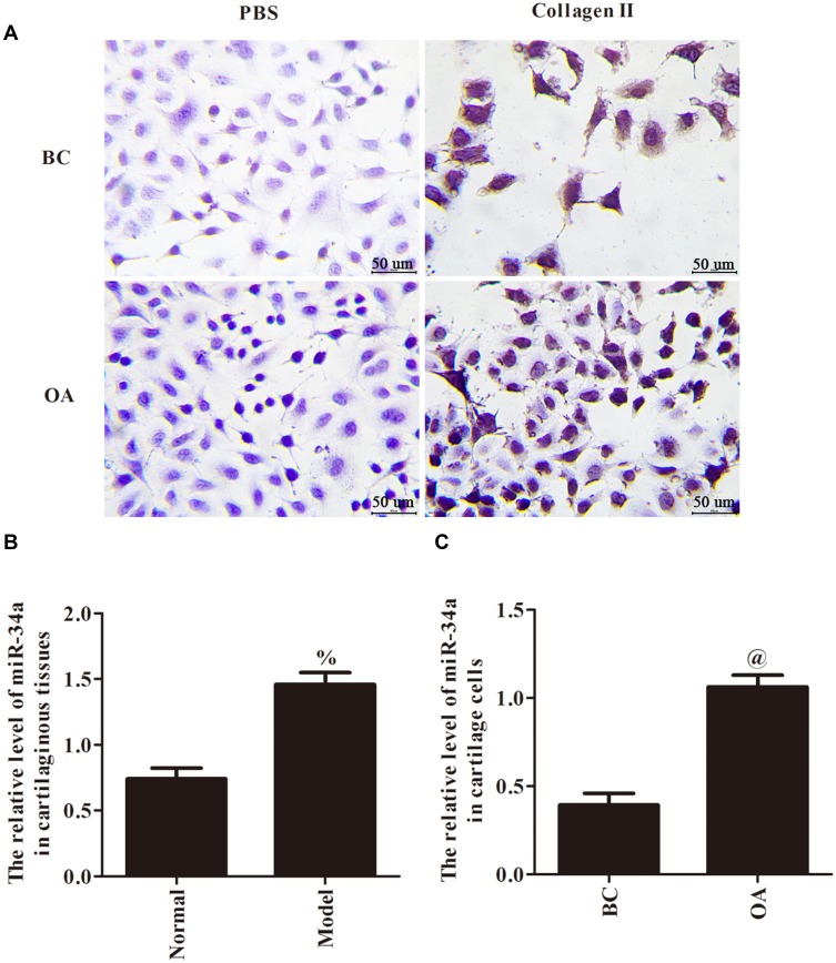
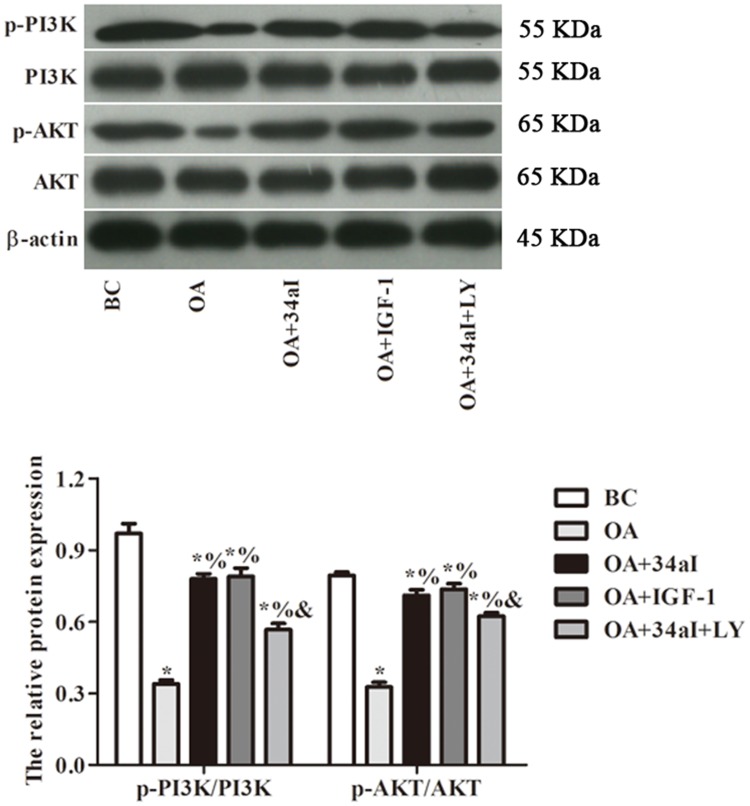
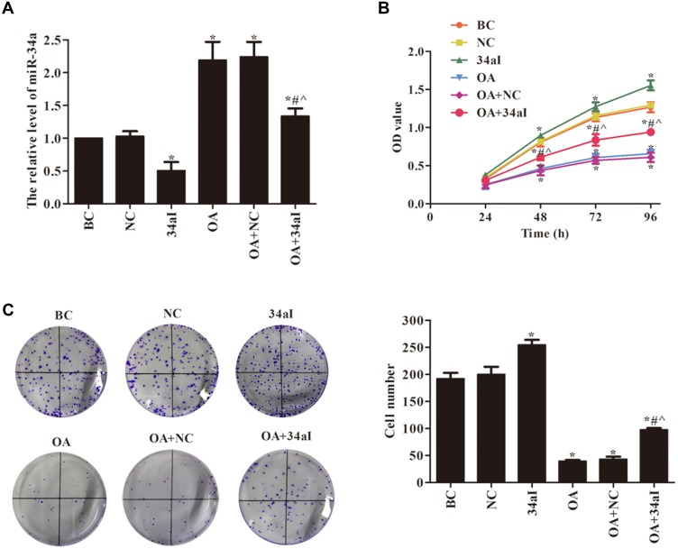
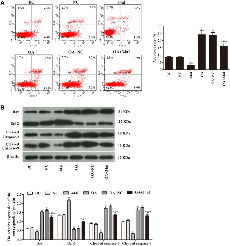
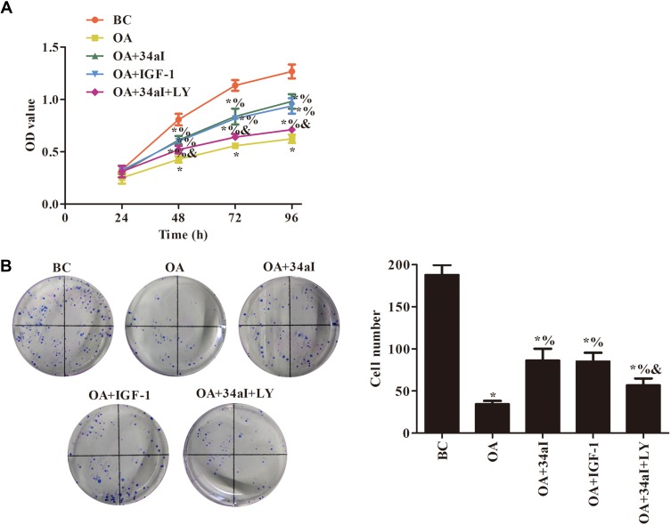
Rat model of osteoarthritis was constructed and knee joint cartilage cells were isolated in vitro. Immunocytochemical staining was used for identification. qRT-PCR was used to detect the expression of miR-34a in cartilaginous tissues and cartilage cells. Cartilage cells were divided into blank control (BC), negative control (NC), miR-34a inhibitor (34aI), osteoarthritis model (OA), osteoarthritis model + negative control (OA + NC) and osteoarthritis model + miR-34a inhibitor (OA + 34aI) groups. Cell proliferation was detected by CCK-8 and colony formation assays. Cell apoptosis was studied by flow cytometry and Western blot. PI3K/AKT-pathway-related proteins were also analyzed by Western blot. To further validate the effect of miR-34a on the PI3K/Akt pathway, the cartilage cells were divided into blank control (BC), osteoarthritis model (OA), osteoarthritis model + miR-34a inhibitor (OA + 34aI), osteoarthritis model + PI3K activator (OA + IGF-1) and osteoarthritis model + miR-34a inhibitor + PI3K inhibitor (OA + 34aI + LY) groups, the experiments above were repeated.
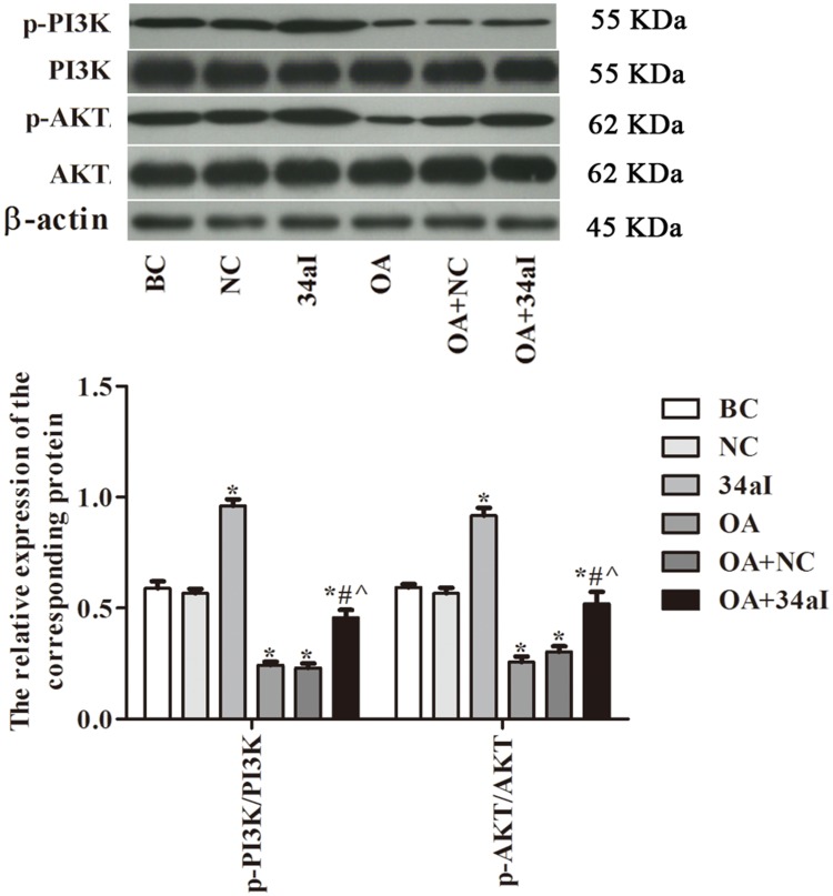
The expression of miR-34a in cartilaginous tissues and cells of osteoarthritis model was significantly higher than that in normal (p < 0.05). After silencing miR-34a gene, the cell proliferation and proteins expression of PI3K/Akt pathway were increased, while the apoptosis rate and expression of apoptosis-related proteins were decreased. Addition of PI3K activator also evidently promoted proliferation and inhibited apoptosis. The protein expression of Bax, Cleaved caspase-3 and Cleaved caspase-9 were dramatically decreased, while the ratios of p-PI3K/PI3K and p-Akt/Akt were increased in OA + IGF-1 group.
Downregulation of miR-34a regulated proliferation and apoptosis of cartilage cells by activating PI3K/Akt pathway, providing a potential therapeutic approach for the treatment of osteoarthritis.
阐明 miR-34a 在大鼠骨关节炎软骨细胞中的表达和功能,并进一步探讨其机制。
构建大鼠骨关节炎模型,体外分离膝关节软骨细胞。免疫细胞化学染色鉴定。qRT-PCR 检测软骨组织和软骨细胞中 miR-34a 的表达。将软骨细胞分为空白对照组(BC)、阴性对照组(NC)、miR-34a 抑制剂组(34aI)、骨关节炎模型组(OA)、骨关节炎模型+阴性对照组(OA+NC)和骨关节炎模型+miR-34a 抑制剂组(OA+34aI)。CCK-8 和集落形成实验检测细胞增殖。流式细胞术和 Western blot 研究细胞凋亡。Western blot 分析 PI3K/AKT 通路相关蛋白。为了进一步验证 miR-34a 对 PI3K/Akt 通路的影响,将软骨细胞分为空白对照组(BC)、骨关节炎模型组(OA)、骨关节炎模型+miR-34a 抑制剂组(OA+34aI)、骨关节炎模型+PI3K 激活剂组(OA+IGF-1)和骨关节炎模型+miR-34a 抑制剂+PI3K 抑制剂组(OA+34aI+LY),重复上述实验。
骨关节炎模型软骨组织和细胞中 miR-34a 的表达明显高于正常组(p<0.05)。沉默 miR-34a 基因后,PI3K/Akt 通路的细胞增殖和蛋白表达增加,而细胞凋亡率和凋亡相关蛋白表达减少。加入 PI3K 激活剂也明显促进增殖,抑制凋亡。OA+IGF-1 组 Bax、Cleaved caspase-3 和 Cleaved caspase-9 的蛋白表达明显降低,p-PI3K/PI3K 和 p-Akt/Akt 的比值增加。
下调 miR-34a 通过激活 PI3K/Akt 通路调节软骨细胞的增殖和凋亡,为骨关节炎的治疗提供了一种潜在的治疗方法。