Yogarajah M, Powell H W R, Parker G J M, Alexander D C, Thompson P J, Symms M R, Boulby P, Wheeler-Kingshott C A, Barker G J, Koepp M J, Duncan J S
Department of Clinical and Experimental Epilepsy, Institute of Neurology, University College London, UK.
Neuroimage. 2008 May 1;40(4):1755-64. doi: 10.1016/j.neuroimage.2007.12.046. Epub 2008 Jan 10.
Temporal lobe epilepsy (TLE) is associated with disrupted memory function. The structural changes underlying this memory impairment have not been demonstrated previously with tractography.
We performed a tractography analysis of diffusion magnetic resonance imaging scans in 18 patients with unilateral TLE undergoing presurgical evaluation, and in 10 healthy controls. A seed region in the anterior parahippocampal gyrus was selected from which to trace the white matter connections of the medial temporal lobe. A correlation analysis was carried out between volume and mean fractional anisotropy (FA) of the connections, and pre-operative material specific memory performance.
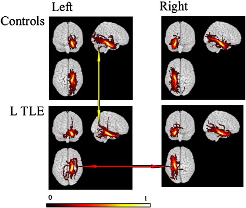
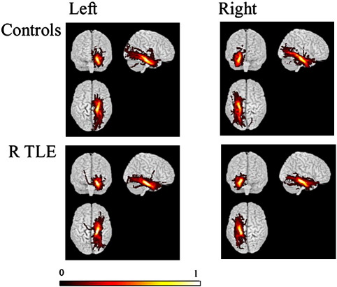
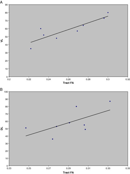
There was no significant difference between the left and right sided connections in controls. In the left TLE patients, the connected regions ipsilateral to the epileptogenic region were found to be significantly reduced in volume and mean FA compared with the contralateral region, and left-sided connections in control subjects. Significant correlations were found in left TLE patients between left and right FA, and verbal and non-verbal memory respectively.
Tractography demonstrated the alteration of white matter pathways that may underlie impaired memory function in TLE. A detailed knowledge of the integrity of these connections may be useful in predicting memory decline in chronic temporal lobe epilepsy.
颞叶癫痫(TLE)与记忆功能紊乱有关。以往的纤维束成像尚未证实这种记忆障碍背后的结构变化。
我们对18例接受术前评估的单侧TLE患者和10名健康对照者的扩散磁共振成像扫描进行了纤维束成像分析。从前海马旁回选择一个种子区域,从中追踪内侧颞叶的白质连接。对连接的体积和平均分数各向异性(FA)与术前特定材料的记忆表现进行相关性分析。
对照组左右侧连接无显著差异。在左侧TLE患者中,与癫痫发作区域同侧的连接区域与对侧区域及对照组左侧连接相比,体积和平均FA显著减小。在左侧TLE患者中,分别在左右FA与言语和非言语记忆之间发现显著相关性。
纤维束成像显示了白质通路的改变,这可能是TLE记忆功能受损的基础。详细了解这些连接的完整性可能有助于预测慢性颞叶癫痫患者的记忆衰退。