Drummond Micah J, Fujita Satoshi, Abe Takashi, Dreyer Hans C, Volpi Elena, Rasmussen Blake B
Department of Physical Therapy, University of Texas Medical Branch, Galveston, TX, USA.
Med Sci Sports Exerc. 2008 Apr;40(4):691-8. doi: 10.1249/MSS.0b013e318160ff84.
Blood flow restriction in combination with low-intensity resistance exercise (REFR) increases skeletal muscle size to a similar extent as compared with traditional high-intensity resistance exercise training. However, there are limited data describing the molecular adaptations that occur after REFR.
To determine whether hypoxia inducible factor-1 alpha (HIF-1alpha) and REDD1 mRNA are expressed differently in REFR compared with low-intensity resistance exercise with no blood flow restriction (CONTROL). Secondly, to determine whether low-intensity resistance exercise is able to induce changes in mRNA expression of several anabolic and catabolic genes as typically seen with high-intensity resistance exercise.
Six subjects were studied at baseline and 3 h after a bout of leg resistance exercise (20% 1RM) in REFR and CONTROL subjects. Each subject participated in both groups, with 3 wk separating each visit. Muscle biopsy samples were analyzed for mRNA expression, using qRT-PCR.
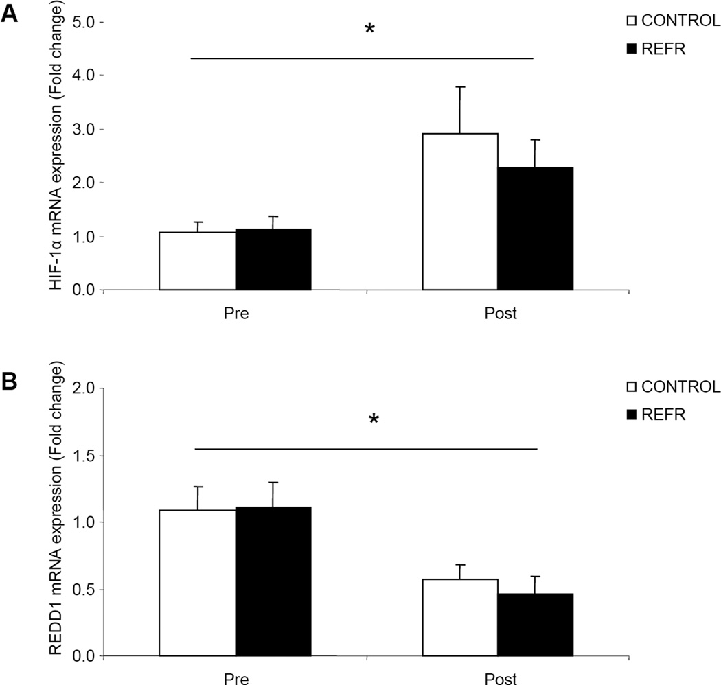
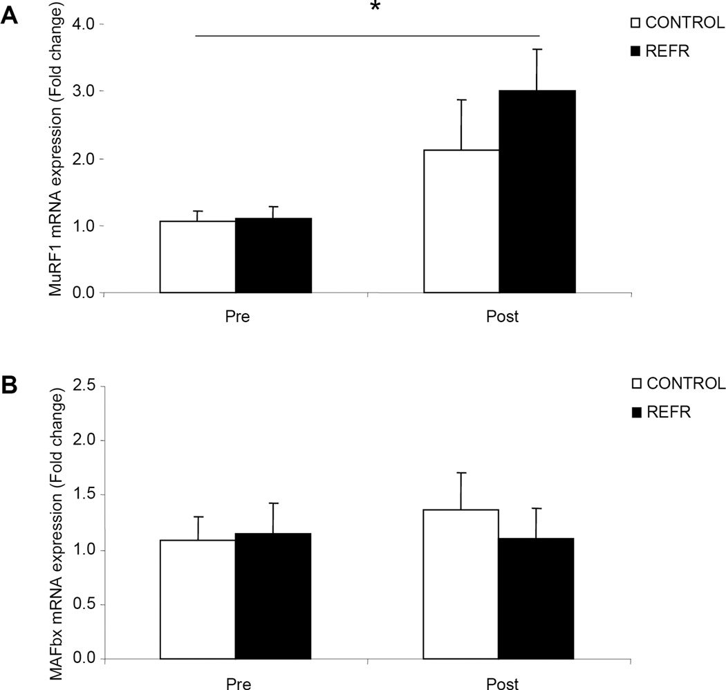
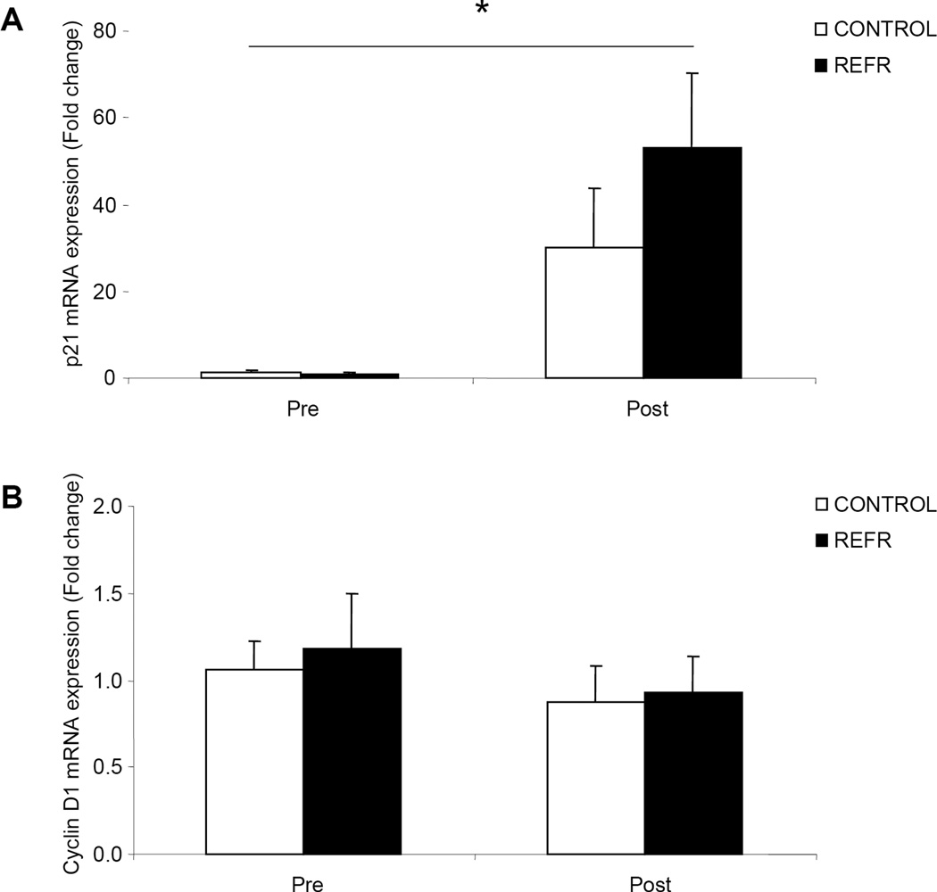
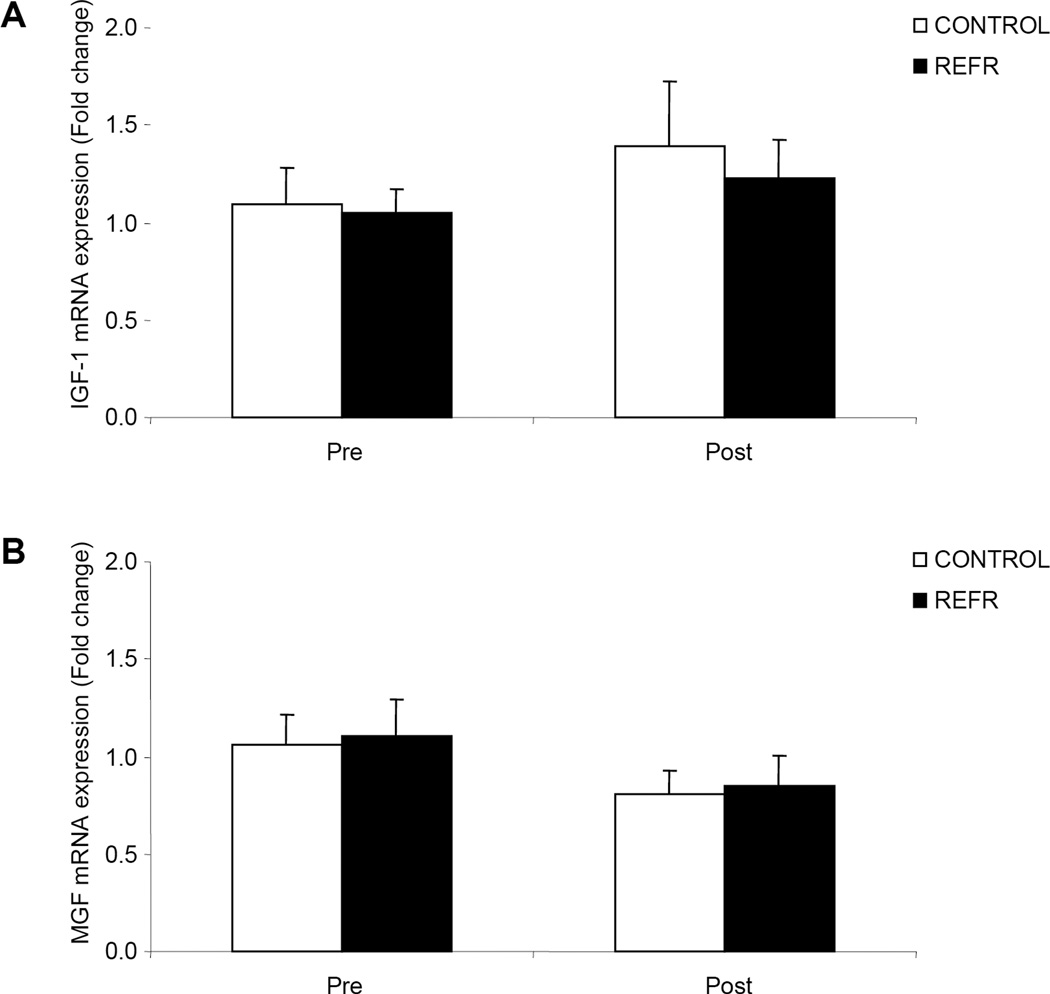
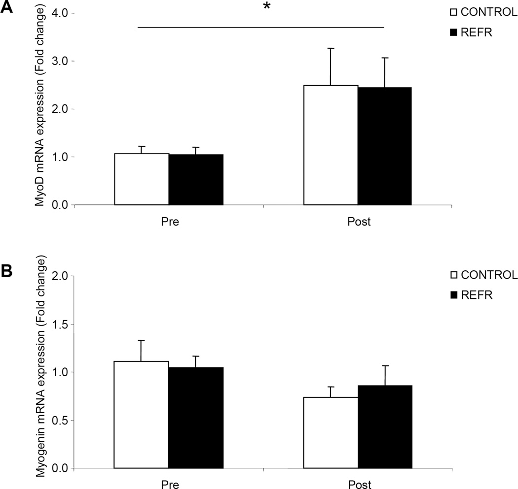
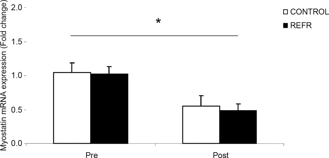
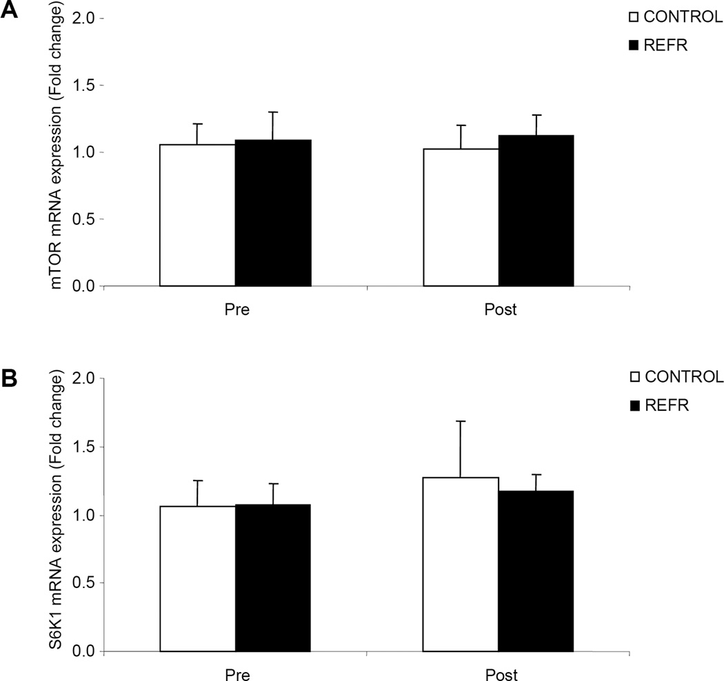
Our primary finding was that there were no differences between CONTROL and REFR for any of the selected genes at 3 h after exercise (P > 0.05). However, low-intensity resistance exercise increased HIF-1alpha, p21, MyoD, and muscle RING finger 1 (MuRF1) mRNA expression and decreased REDD1 and myostatin mRNA expression in both groups (P < 0.05).
Low-intensity resistance exercise can alter skeletal muscle mRNA expression of several genes associated with muscle growth and remodeling, such as REDD1, HIF-1alpha, MyoD, MuRF1, and myostatin. Further, the results from REFR and CONTROL were similar, indicating that the changes in early postexercise gene expression were attributable to the low-intensity resistance exercise bout, and not blood flow restriction.
与传统高强度抗阻训练相比,血流限制联合低强度抗阻运动(REFR)可使骨骼肌大小增加至相似程度。然而,关于REFR后发生的分子适应性变化的数据有限。
确定与无血流限制的低强度抗阻运动(对照组)相比,低氧诱导因子-1α(HIF-1α)和REDD1 mRNA在REFR中是否有不同表达。其次,确定低强度抗阻运动是否能够像高强度抗阻运动那样诱导几种合成代谢和分解代谢基因的mRNA表达发生变化。
对6名受试者在基线时以及在REFR组和对照组进行一组腿部抗阻运动(20% 1RM)后3小时进行研究。每名受试者参加两组实验,每次就诊间隔3周。使用qRT-PCR分析肌肉活检样本的mRNA表达。
我们的主要发现是,运动后3小时,所选基因在对照组和REFR组之间均无差异(P>0.05)。然而,低强度抗阻运动使两组中的HIF-1α、p21、MyoD和肌肉环形指蛋白1(MuRF1)mRNA表达增加,REDD1和肌生成抑制素mRNA表达降低(P<0.05)。
低强度抗阻运动可改变骨骼肌中与肌肉生长和重塑相关的几种基因的mRNA表达,如REDD1、HIF-1α、MyoD、MuRF1和肌生成抑制素。此外,REFR组和对照组的结果相似,表明运动后早期基因表达的变化归因于低强度抗阻运动,而非血流限制。