• 文献检索
  • 文档翻译
  • 深度研究
  • 学术资讯
  • Suppr Zotero 插件Zotero 插件
  • 邀请有礼
  • 套餐&价格
  • 历史记录
应用&插件
Suppr Zotero 插件Zotero 插件浏览器插件Mac 客户端Windows 客户端微信小程序
定价
高级版会员购买积分包购买API积分包
服务
文献检索文档翻译深度研究API 文档MCP 服务
关于我们
关于 Suppr公司介绍联系我们用户协议隐私条款
关注我们

Suppr 超能文献

核心技术专利:CN118964589B侵权必究
粤ICP备2023148730 号-1Suppr @ 2026

文献检索

告别复杂PubMed语法,用中文像聊天一样搜索,搜遍4000万医学文献。AI智能推荐,让科研检索更轻松。

立即免费搜索

文件翻译

保留排版,准确专业,支持PDF/Word/PPT等文件格式,支持 12+语言互译。

免费翻译文档

深度研究

AI帮你快速写综述,25分钟生成高质量综述,智能提取关键信息,辅助科研写作。

立即免费体验

与特定通气的氙CT测量结果相比,基于配准的局部肺组织扩张估计。

Registration-based estimates of local lung tissue expansion compared to xenon CT measures of specific ventilation.

作者信息

Reinhardt Joseph M, Ding Kai, Cao Kunlin, Christensen Gary E, Hoffman Eric A, Bodas Shalmali V

机构信息

Department of Biomedical Engineering, University of Iowa, Iowa City, IA 52242, USA.

出版信息

Med Image Anal. 2008 Dec;12(6):752-63. doi: 10.1016/j.media.2008.03.007. Epub 2008 Apr 12.

DOI:10.1016/j.media.2008.03.007
PMID:18501665
原文链接:https://pmc.ncbi.nlm.nih.gov/articles/PMC2692217/
Abstract

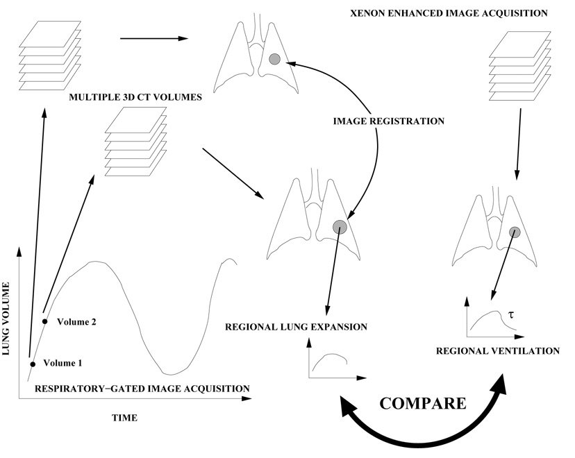
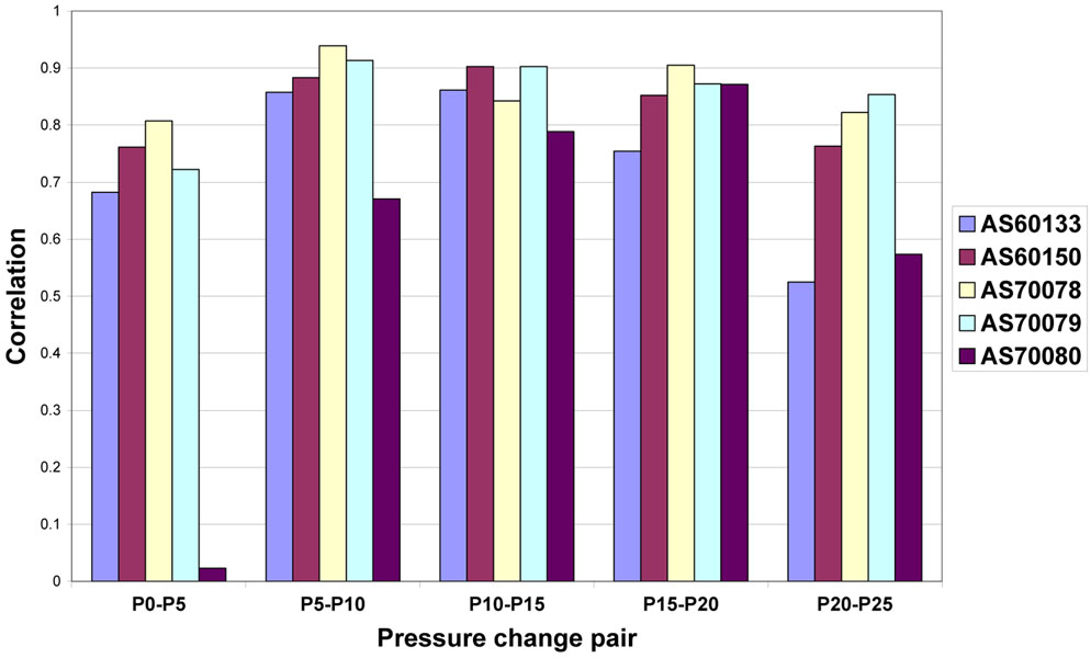
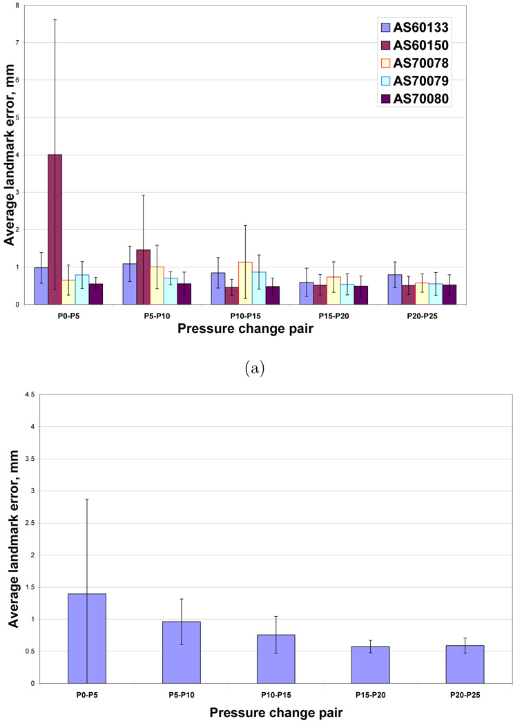
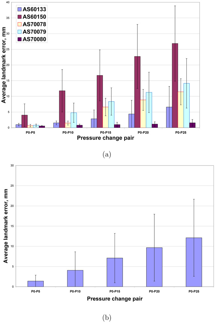
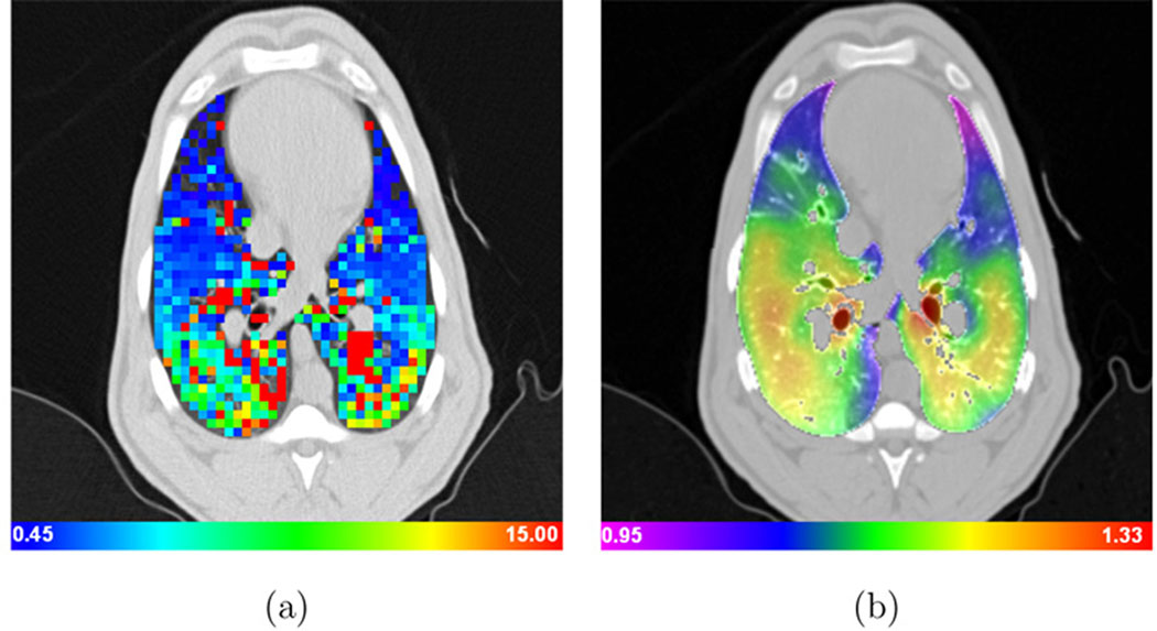
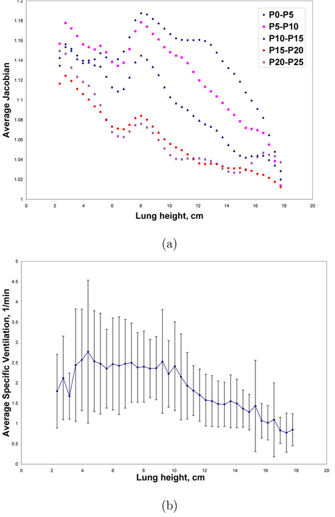
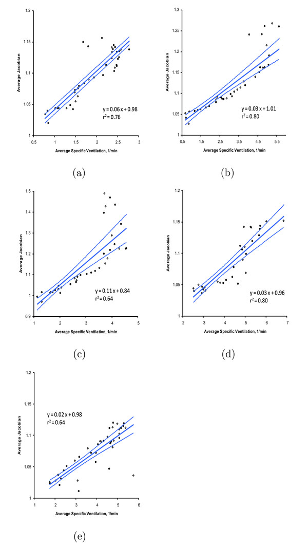
The main function of the respiratory system is gas exchange. Since many disease or injury conditions can cause biomechanical or material property changes that can alter lung function, there is a great interest in measuring regional lung ventilation and regional specific volume change. We describe a registration-based technique for estimating local lung expansion from multiple respiratory-gated CT images of the thorax. The degree of regional lung expansion is measured using the Jacobian (a function of local partial derivatives) of the registration displacement field, which we show is directly related to specific volume change. We compare the ventral-dorsal patterns of lung expansion estimated across five pressure changes to a xenon CT based measure of specific ventilation in five anesthetized sheep studied in the supine orientation. Using 3D image registration to match images acquired at 10 cm H(2)O and 15 cm H(2)O airway pressures gave the best match between the average Jacobian and the xenon CT specific ventilation (linear regression, average r(2)=0.73).

摘要

呼吸系统的主要功能是气体交换。由于许多疾病或损伤情况会导致生物力学或材料特性的变化,进而改变肺功能,因此人们对测量局部肺通气和局部比容变化有着浓厚的兴趣。我们描述了一种基于配准的技术,用于从胸部的多个呼吸门控CT图像估计局部肺扩张。使用配准位移场的雅可比行列式(局部偏导数的函数)来测量局部肺扩张程度,我们证明它与比容变化直接相关。我们将在仰卧位研究的五只麻醉绵羊中,在五个压力变化下估计的肺扩张腹背模式与基于氙CT的比通气量测量值进行了比较。使用三维图像配准来匹配在气道压力为10 cm H₂O和15 cm H₂O时采集的图像,平均雅可比行列式与氙CT比通气量之间的匹配度最佳(线性回归,平均r² = 0.73)。

https://cdn.ncbi.nlm.nih.gov/pmc/blobs/26d6/2692217/ae20fb0210a9/nihms78809f13.jpg
https://cdn.ncbi.nlm.nih.gov/pmc/blobs/26d6/2692217/1ac338cc4cb1/nihms78809f1.jpg
https://cdn.ncbi.nlm.nih.gov/pmc/blobs/26d6/2692217/b31f8f181517/nihms78809f2.jpg
https://cdn.ncbi.nlm.nih.gov/pmc/blobs/26d6/2692217/476e48284956/nihms78809f3.jpg
https://cdn.ncbi.nlm.nih.gov/pmc/blobs/26d6/2692217/beccf1afa0f1/nihms78809f4.jpg
https://cdn.ncbi.nlm.nih.gov/pmc/blobs/26d6/2692217/3193a557c85b/nihms78809f5.jpg
https://cdn.ncbi.nlm.nih.gov/pmc/blobs/26d6/2692217/216c480efde6/nihms78809f6.jpg
https://cdn.ncbi.nlm.nih.gov/pmc/blobs/26d6/2692217/1cc767af7fb9/nihms78809f7.jpg
https://cdn.ncbi.nlm.nih.gov/pmc/blobs/26d6/2692217/e76cb1d4b0c5/nihms78809f8.jpg
https://cdn.ncbi.nlm.nih.gov/pmc/blobs/26d6/2692217/d2bdf08adccf/nihms78809f9.jpg
https://cdn.ncbi.nlm.nih.gov/pmc/blobs/26d6/2692217/5e77c7b02705/nihms78809f10.jpg
https://cdn.ncbi.nlm.nih.gov/pmc/blobs/26d6/2692217/e4011a3e9786/nihms78809f11.jpg
https://cdn.ncbi.nlm.nih.gov/pmc/blobs/26d6/2692217/87340340008a/nihms78809f12.jpg
https://cdn.ncbi.nlm.nih.gov/pmc/blobs/26d6/2692217/ae20fb0210a9/nihms78809f13.jpg

相似文献

1
Registration-based estimates of local lung tissue expansion compared to xenon CT measures of specific ventilation.与特定通气的氙CT测量结果相比,基于配准的局部肺组织扩张估计。
Med Image Anal. 2008 Dec;12(6):752-63. doi: 10.1016/j.media.2008.03.007. Epub 2008 Apr 12.
2
Registration-derived estimates of local lung expansion as surrogates for regional ventilation.通过配准得出的局部肺扩张估计值作为区域通气的替代指标。
Inf Process Med Imaging. 2007;20:763-74. doi: 10.1007/978-3-540-73273-0_63.
3
Reproducibility of intensity-based estimates of lung ventilation.基于强度的肺通气估计的可重复性。
Med Phys. 2013 Jun;40(6):063504. doi: 10.1118/1.4805106.
4
Comparison of image registration based measures of regional lung ventilation from dynamic spiral CT with Xe-CT.基于动态螺旋CT的区域肺通气图像配准测量与氙CT的比较。
Med Phys. 2012 Aug;39(8):5084-98. doi: 10.1118/1.4736808.
5
Dynamic ventilation imaging from four-dimensional computed tomography.基于四维计算机断层扫描的动态通气成像。
Phys Med Biol. 2006 Feb 21;51(4):777-91. doi: 10.1088/0031-9155/51/4/002. Epub 2006 Jan 25.
6
Tracking lung tissue motion and expansion/compression with inverse consistent image registration and spirometry.使用反向一致图像配准和肺活量测定法追踪肺组织运动及扩张/压缩情况。
Med Phys. 2007 Jun;34(6):2155-63. doi: 10.1118/1.2731029.
7
4D-CT motion estimation using deformable image registration and 5D respiratory motion modeling.使用可变形图像配准的4D-CT运动估计和5D呼吸运动建模。
Med Phys. 2008 Oct;35(10):4577-90. doi: 10.1118/1.2977828.
8
Subtraction lung image for evaluating pulmonary ventilation in xenon-enhanced CT.用于评估氙增强 CT 中肺通气的减影肺图像。
Med Phys. 2010 Aug;37(8):4464-74. doi: 10.1118/1.3464490.
9
Lung lobar slippage assessed with the aid of image registration.借助图像配准评估肺叶滑动。
Med Image Comput Comput Assist Interv. 2010;13(Pt 2):578-85. doi: 10.1007/978-3-642-15745-5_71.
10
Ray-tracing based registration for HRCT images of the lungs.基于光线追踪的肺部高分辨率CT图像配准
Med Image Comput Comput Assist Interv. 2006;9(Pt 2):670-7.

引用本文的文献

1
Weekly changes in ventilation response for photon and proton lung cancer patients during radiotherapy.肺癌患者在放疗期间光子和质子治疗时通气反应的每周变化。
medRxiv. 2025 Aug 29:2025.08.28.25334578. doi: 10.1101/2025.08.28.25334578.
2
[Research progress on predicting radiation pneumonia based on four-dimensional computed tomography ventilation imaging in lung cancer radiotherapy].[基于四维计算机断层扫描通气成像预测肺癌放疗中放射性肺炎的研究进展]
Sheng Wu Yi Xue Gong Cheng Xue Za Zhi. 2025 Aug 25;42(4):863-870. doi: 10.7507/1001-5515.202405008.
3
Quantifying changes in ventilation for photon- and proton-treated patients according to dose and pneumonitis development.

本文引用的文献

1
Hyperpolarized (129)Xe MRI: a viable functional lung imaging modality?超极化(129)Xe磁共振成像:一种可行的肺部功能成像方式?
Eur J Radiol. 2007 Dec;64(3):335-44. doi: 10.1016/j.ejrad.2007.08.008. Epub 2007 Sep 24.
2
Tracking lung tissue motion and expansion/compression with inverse consistent image registration and spirometry.使用反向一致图像配准和肺活量测定法追踪肺组织运动及扩张/压缩情况。
Med Phys. 2007 Jun;34(6):2155-63. doi: 10.1118/1.2731029.
3
Registration-derived estimates of local lung expansion as surrogates for regional ventilation.
根据剂量和肺炎发展情况,对接受光子和质子治疗的患者的通气变化进行量化。
medRxiv. 2025 Aug 19:2025.03.06.25323515. doi: 10.1101/2025.03.06.25323515.
4
Assessing the consistency of CT-based ventilation imaging under noise reduction processing with simulated quantum noise using a nonrigid alveoli phantom.使用非刚性肺泡模型评估基于CT的通气成像在降噪处理下与模拟量子噪声的一致性。
Front Radiol. 2025 Jul 9;5:1567267. doi: 10.3389/fradi.2025.1567267. eCollection 2025.
5
Forward Computational Modeling of Respiratory Airflow.呼吸气流的正向计算建模
Appl Sci (Basel). 2024 Dec 2;14(24). doi: 10.3390/app142411591. Epub 2024 Dec 12.
6
Use of quantitative CT chest imaging to derive and assess a radiographic phenotype of deployment-related constrictive bronchiolitis.使用胸部定量CT成像来推导和评估与部署相关的缩窄性细支气管炎的影像学表型。
Respir Res. 2025 May 21;26(1):195. doi: 10.1186/s12931-025-03269-8.
7
More accessible functional lung imaging: non-contrast CT-ventilation demonstrates strong association and agreement with PET-ventilation.更易获得的功能性肺成像:非增强CT通气与PET通气显示出很强的相关性和一致性。
Respir Res. 2025 Apr 26;26(1):163. doi: 10.1186/s12931-025-03245-2.
8
PixelPrint4D: A 3D Printing Method of Fabricating Patient-Specific Deformable CT Phantoms for Respiratory Motion Applications.PixelPrint4D:一种用于呼吸运动应用的定制可变形CT体模的3D打印方法。
Invest Radiol. 2025 Apr 2. doi: 10.1097/RLI.0000000000001182.
9
Hybrid method for estimating lung ventilation from CT by combining intensity and motion information.通过结合强度和运动信息从CT估计肺通气的混合方法。
Med Phys. 2025 Jun;52(6):4528-4539. doi: 10.1002/mp.17787. Epub 2025 Mar 30.
10
TLRN: Temporal Latent Residual Networks For Large Deformation Image Registration.TLRN:用于大变形图像配准的时间潜在残差网络
Med Image Comput Comput Assist Interv. 2024 Oct;15002:728-738. doi: 10.1007/978-3-031-72069-7_68. Epub 2024 Oct 4.
通过配准得出的局部肺扩张估计值作为区域通气的替代指标。
Inf Process Med Imaging. 2007;20:763-74. doi: 10.1007/978-3-540-73273-0_63.
4
Effect of low-xenon and krypton supplementation on signal/noise of regional CT-based ventilation measurements.低氙气和氪气补充对基于CT的区域通气测量信号/噪声的影响。
J Appl Physiol (1985). 2007 Apr;102(4):1535-44. doi: 10.1152/japplphysiol.01235.2005. Epub 2006 Nov 22.
5
Assessment of lung development using hyperpolarized helium-3 diffusion MR imaging.使用超极化氦-3扩散磁共振成像评估肺发育
J Magn Reson Imaging. 2006 Dec;24(6):1277-83. doi: 10.1002/jmri.20723.
6
Dynamic ventilation imaging from four-dimensional computed tomography.基于四维计算机断层扫描的动态通气成像。
Phys Med Biol. 2006 Feb 21;51(4):777-91. doi: 10.1088/0031-9155/51/4/002. Epub 2006 Jan 25.
7
Differences in regional wash-in and wash-out time constants for xenon-CT ventilation studies.氙CT通气研究中区域肺内气体充盈和排空时间常数的差异。
Respir Physiol Neurobiol. 2005 Aug 25;148(1-2):65-83. doi: 10.1016/j.resp.2005.06.003.
8
Quantification of regional ventilation from treatment planning CT.通过治疗计划CT进行区域通气的量化
Int J Radiat Oncol Biol Phys. 2005 Jul 1;62(3):630-4. doi: 10.1016/j.ijrobp.2005.03.023.
9
Towards a model of lung biomechanics: pulmonary kinematics via registration of serial lung images.迈向肺生物力学模型:通过连续肺图像配准实现肺运动学
Med Image Anal. 2005 Dec;9(6):524-37. doi: 10.1016/j.media.2005.04.002.
10
Combined helium-3/proton magnetic resonance imaging measurement of ventilated lung volumes in smokers compared to never-smokers.与从不吸烟者相比,吸烟者通气肺容积的氦-3/质子联合磁共振成像测量
J Magn Reson Imaging. 2005 Apr;21(4):365-9. doi: 10.1002/jmri.20290.