Noworolski Susan Moyher, Vigneron Daniel B, Chen Albert P, Kurhanewicz John
Department of Radiology and Biomedical Imaging, The University of California, San Francisco, San Francisco, CA, USA.
Magn Reson Imaging. 2008 Oct;26(8):1071-80. doi: 10.1016/j.mri.2008.01.033. Epub 2008 May 27.
To compare peak enhancement (PE), determined from dynamic contrast-enhanced (DCE) magnetic resonance imaging (MRI) and the magnetic resonance (MR) directionally-averaged apparent diffusion coefficient (
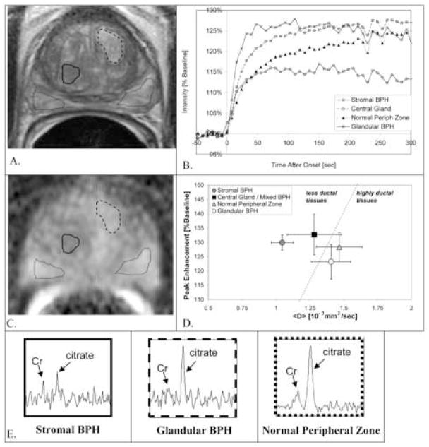
MRI, MR spectroscopic imaging, DCE MRI and MR diffusion were evaluated in 17 untreated subjects with suspected or proven prostate cancer. PE and
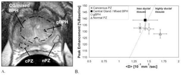
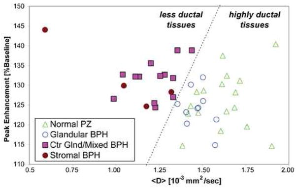
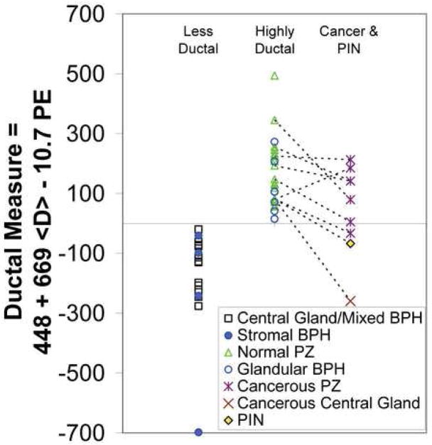
The glandular-ductal tissues had lower PE [125+/-6.4 (% baseline)] and higher
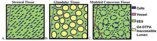
The very different MR results in the glandular-ductal versus stromal-low ductal tissues suggest that these tissues have different underlying structure. These results support the hypothesis that Gd-DTPA does not enter healthy prostatic glands or ducts. This may explain the higher PE and lower
比较动态对比增强(DCE)磁共振成像(MRI)测定的峰值增强(PE)以及前列腺腺性组织与基质组织中的磁共振(MR)方向平均表观扩散系数(
对17名未经治疗的疑似或确诊前列腺癌患者进行MRI、磁共振波谱成像、DCE MRI和MR扩散评估。比较腺管组织[正常外周带和腺性良性前列腺增生(BPH)]与基质 - 低导管组织(中央腺体/混合性BPH和基质性BPH)中的PE和
腺管组织的PE[125±6.4(%基线)]低于基质 - 低导管组织[PE = 132±5.5(%基线)(P <.0008)],而
腺管组织与基质 - 低导管组织中截然不同的MR结果表明这些组织具有不同的基础结构。这些结果支持Gd - DTPA不进入健康前列腺腺体或导管这一假设。这可能解释了先前报道的前列腺癌与健康组织相比更高的PE和更低的