Jin Jing-Ji, Kim Hong-Duck, Maxwell J Adam, Li Ling, Fukuchi Ken-Ichiro
Department of Cancer Biology and Pharmacology, University of Illinois College of Medicine at Peoria, Box 1649, Peoria, IL 61656, USA.
J Neuroinflammation. 2008 May 29;5:23. doi: 10.1186/1742-2094-5-23.
Abeta deposits in the brains of patients with Alzheimer's disease (AD) are closely associated with innate immune responses such as activated microglia and increased cytokines. Accumulating evidence supports the hypothesis that innate immune/inflammatory responses play a pivotal role in the pathogenesis of AD: either beneficial or harmful effects on the AD progression. The molecular mechanisms by which the innate immune system modulates the AD progression are not well understood. Toll-like receptors (TLRs) are first-line molecules for initiating the innate immune responses. When activated through TLR signaling, microglia respond to pathogens and damaged host cells by secreting chemokines and cytokines and express the co-stimulatory molecules needed for protective immune responses to pathogens and efficient clearance of damaged tissues. We previously demonstrated that an AD mouse model homozygous for a destructive mutation of TLR4 has increases in diffuse and fibrillar Abeta deposits as well as buffer-soluble and insoluble Abeta in the brain as compared with a TLR4 wild-type AD mouse model. Here, we investigated the roles of TLR4 in Abeta-induced upregulation of cytokines and chemokines, Abeta-induced activation of microglia and astrocytes and Abeta-induced immigration of leukocytes.
Using the same model, levels of cytokines and chemokines in the brain were determined by multiplex cytokine/chemokine array. Activation of microglia and astrocytes and immigration of leukocytes were determined by immunoblotting and immunohistochemistry followed by densitometry and morphometry, respectively.
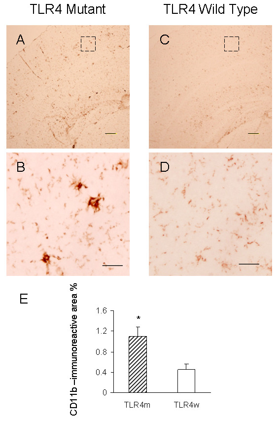
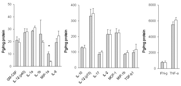
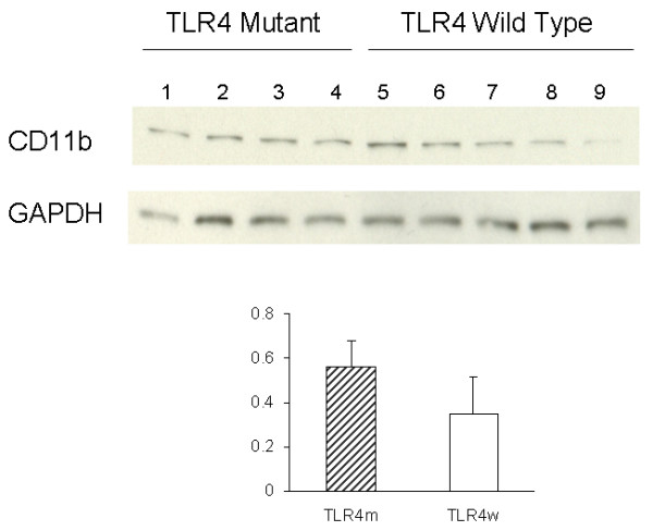
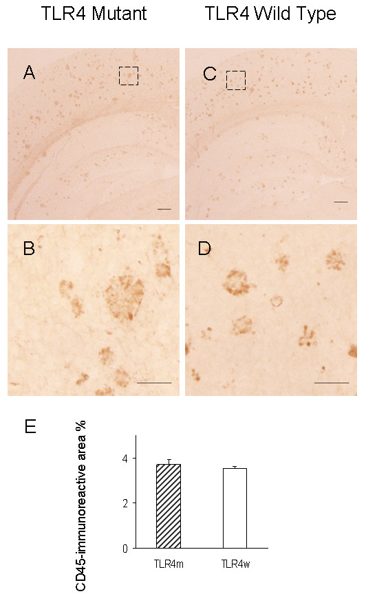
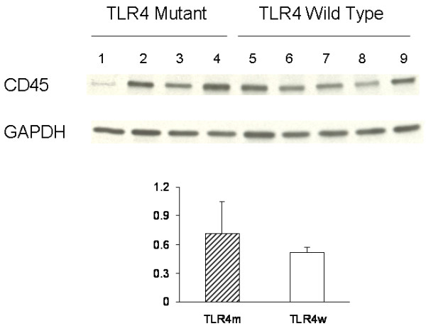
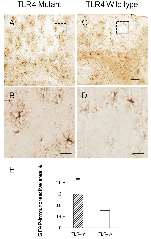
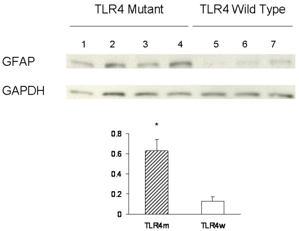
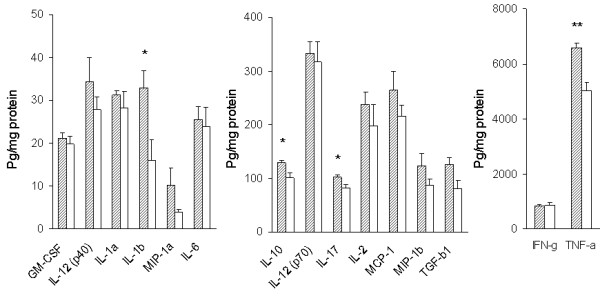
Levels of tumor necrosis factor (TNF)-alpha, interleukin (IL)-1beta, IL-10 and IL-17 in the brains of TLR4 wild-type AD mice were significantly higher than those in TLR4 wild-type non-transgenic littermates. Such increases in cytokines were not found in TLR4 mutant AD mice as compared with TLR4 mutant non-transgenic littermates. Although expression levels of CD11b (a microglia marker) and GFAP (a reactive astrocyte marker) in the brains of TLR4 mutant AD mice were higher than those in TLR4 wild type AD mice, no difference was found in levels of CD45 (common leukocyte antigen).
This is the first demonstration of TLR4-dependent upregulation of cytokines in an AD mouse model. Our results suggest that TLR4 signaling is involved in AD progression and that TLR4 signaling can be a new therapeutic target for AD.
阿尔茨海默病(AD)患者大脑中的淀粉样蛋白β(Aβ)沉积与先天性免疫反应密切相关,如小胶质细胞活化和细胞因子增加。越来越多的证据支持这样一种假说,即先天性免疫/炎症反应在AD发病机制中起关键作用:对AD进展既有有益影响,也有有害影响。先天性免疫系统调节AD进展的分子机制尚不清楚。Toll样受体(TLR)是启动先天性免疫反应的一线分子。当通过TLR信号激活时,小胶质细胞通过分泌趋化因子和细胞因子对病原体和受损宿主细胞作出反应,并表达对病原体进行保护性免疫反应和有效清除受损组织所需的共刺激分子。我们之前证明,与TLR4野生型AD小鼠模型相比,TLR4发生破坏性突变的纯合AD小鼠模型大脑中弥漫性和纤维状Aβ沉积以及缓冲液可溶性和不溶性Aβ均有所增加。在此,我们研究了TLR4在Aβ诱导的细胞因子和趋化因子上调、Aβ诱导的小胶质细胞和星形胶质细胞活化以及Aβ诱导的白细胞迁移中的作用。
使用相同模型,通过多重细胞因子/趋化因子阵列测定大脑中细胞因子和趋化因子的水平。分别通过免疫印迹和免疫组织化学,随后进行光密度测定和形态测定,来确定小胶质细胞和星形胶质细胞的活化以及白细胞的迁移。
TLR4野生型AD小鼠大脑中肿瘤坏死因子(TNF)-α、白细胞介素(IL)-1β、IL-10和IL-17的水平显著高于TLR4野生型非转基因同窝小鼠。与TLR4突变型非转基因同窝小鼠相比,TLR4突变型AD小鼠未发现细胞因子有此类增加。尽管TLR4突变型AD小鼠大脑中CD11b(小胶质细胞标志物)和GFAP(反应性星形胶质细胞标志物)的表达水平高于TLR4野生型AD小鼠,但CD45(常见白细胞抗原)水平未发现差异。
这是首次在AD小鼠模型中证明细胞因子的TLR4依赖性上调。我们的结果表明,TLR4信号传导参与AD进展,并且TLR4信号传导可能成为AD的新治疗靶点。