Rappoport Joshua Z, Heyman Katherine P, Kemal Shahrnaz, Simon Sanford M
Laboratory of Cellular Biophysics, The Rockefeller University, New York, New York, United States of America.
PLoS One. 2008 Jun 11;3(6):e2416. doi: 10.1371/journal.pone.0002416.
Members of the dynamin super-family of GTPases are involved in disparate cellular pathways. Dynamin1 and dynamin2 have been implicated in clathrin-mediated endocytosis. While some models suggest that dynamin functions specifically at the point of vesicle fission, evidence also exists for a role prior to fission during vesicle formation and it is unknown if there is a role for dynamin after vesicle fission. Although dynamin2 is ubiquitously expressed, dynamin1 is restricted to the nervous system. These two structurally similar endocytic accessory proteins have not been studied in cells that endogenously express both.
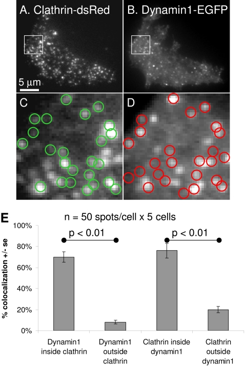
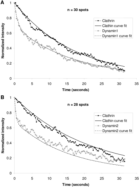
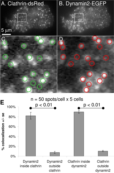
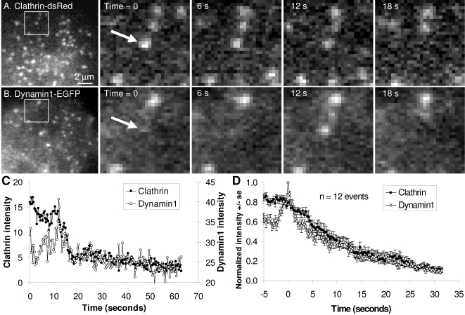
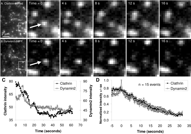
METHODOLOGY/PRINCIPAL FINDINGS: The present study quantitatively assesses the dynamics of dynamin1 and dynamin2 during clathrin-mediated endocytosis in PC12 cells, which endogenously express both proteins. Both dynamin isoforms co-localized with clathrin and showed sharp increases in fluorescence intensity immediately prior to internalization of the nascent clathrin-coated vesicle. The fluorescence intensity of both proteins then decreased with two time constants. The slower time constant closely matched the time constant for the decrease of clathrin intensity and likely represents vesicle movement away from the membrane. The faster rate may reflect release of dynamin at the neck of nascent vesicle following GTP hydrolysis.
CONCLUSIONS/SIGNIFICANCE: This study analyses the role of dynamin in clathrin-mediated endocytosis in a model for cellular neuroscience and these results may provide direct evidence for the existence of two populations of dynamin associated with nascent clathrin-coated vesicles.
GTP酶动力蛋白超家族的成员参与不同的细胞途径。动力蛋白1和动力蛋白2与网格蛋白介导的内吞作用有关。虽然一些模型表明动力蛋白在囊泡分裂点发挥特定作用,但也有证据表明其在囊泡形成过程中分裂前发挥作用,并且囊泡分裂后动力蛋白是否发挥作用尚不清楚。尽管动力蛋白2普遍表达,但动力蛋白1仅限于神经系统。尚未在同时内源性表达这两种蛋白的细胞中对这两种结构相似的内吞辅助蛋白进行研究。
方法/主要发现:本研究定量评估了在PC12细胞(该细胞内源性表达这两种蛋白)的网格蛋白介导的内吞作用过程中动力蛋白1和动力蛋白2的动态变化。两种动力蛋白异构体均与网格蛋白共定位,并在新生网格蛋白包被囊泡内化之前立即显示出荧光强度的急剧增加。然后,两种蛋白的荧光强度以两个时间常数下降。较慢的时间常数与网格蛋白强度下降的时间常数密切匹配,可能代表囊泡从膜上移开。较快的速率可能反映了GTP水解后动力蛋白在新生囊泡颈部的释放。
结论/意义:本研究分析了动力蛋白在细胞神经科学模型中网格蛋白介导的内吞作用中的作用,这些结果可能为与新生网格蛋白包被囊泡相关的两种动力蛋白群体的存在提供直接证据。