Drevets Wayne C, Price Joseph L, Furey Maura L
Section on Neuroimaging in Mood and Anxiety Disorders, National Institute of Mental Health, National Institutes of Health (NIH/NIMH DIRP), 15K North Dr., Room 210, Bethesda, MD 20892, USA.
Brain Struct Funct. 2008 Sep;213(1-2):93-118. doi: 10.1007/s00429-008-0189-x. Epub 2008 Aug 13.
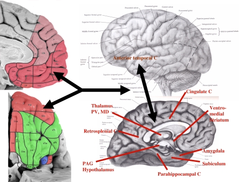
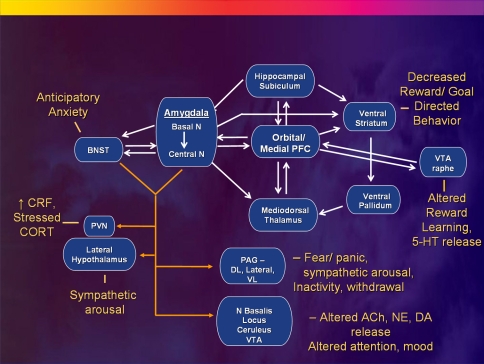
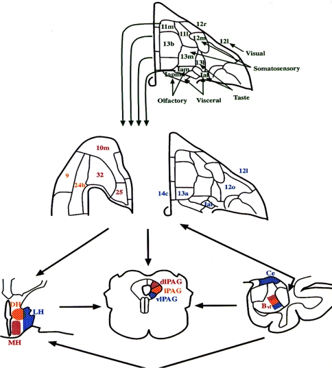
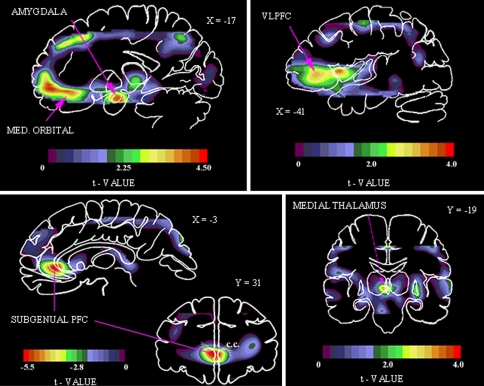
The neural networks that putatively modulate aspects of normal emotional behavior have been implicated in the pathophysiology of mood disorders by converging evidence from neuroimaging, neuropathological and lesion analysis studies. These networks involve the medial prefrontal cortex (MPFC) and closely related areas in the medial and caudolateral orbital cortex (medial prefrontal network), amygdala, hippocampus, and ventromedial parts of the basal ganglia, where alterations in grey matter volume and neurophysiological activity are found in cases with recurrent depressive episodes. Such findings hold major implications for models of the neurocircuits that underlie depression. In particular evidence from lesion analysis studies suggests that the MPFC and related limbic and striato-pallido-thalamic structures organize emotional expression. The MPFC is part of a larger "default system" of cortical areas that include the dorsal PFC, mid- and posterior cingulate cortex, anterior temporal cortex, and entorhinal and parahippocampal cortex, which has been implicated in self-referential functions. Dysfunction within and between structures in this circuit may induce disturbances in emotional behavior and other cognitive aspects of depressive syndromes in humans. Further, because the MPFC and related limbic structures provide forebrain modulation over visceral control structures in the hypothalamus and brainstem, their dysfunction can account for the disturbances in autonomic regulation and neuroendocrine responses that are associated with mood disorders. This paper discusses these systems together with the neurochemical systems that impinge on them and form the basis for most pharmacological therapies.
通过神经影像学、神经病理学和病变分析研究得出的一致证据表明,推测对正常情绪行为各方面进行调节的神经网络与情绪障碍的病理生理学有关。这些神经网络包括内侧前额叶皮质(MPFC)以及内侧和尾外侧眶皮质中密切相关的区域(内侧前额叶网络)、杏仁核、海马体和基底神经节的腹内侧部分,在复发性抑郁发作的病例中发现这些区域的灰质体积和神经生理活动存在改变。这些发现对抑郁症潜在的神经回路模型具有重要意义。特别是来自病变分析研究的证据表明,MPFC以及相关的边缘系统和纹状体 - 苍白球 - 丘脑结构参与情绪表达的组织。MPFC是包括背侧前额叶皮质、扣带前回和后回、颞叶前部皮质以及内嗅皮质和海马旁皮质在内的更大的皮质区域“默认系统”的一部分,该系统与自我参照功能有关。该回路中结构内部和之间的功能障碍可能会导致人类抑郁综合征在情绪行为和其他认知方面出现紊乱。此外,由于MPFC和相关的边缘系统结构对下丘脑和脑干中的内脏控制结构提供前脑调节,它们的功能障碍可以解释与情绪障碍相关的自主神经调节和神经内分泌反应的紊乱。本文将讨论这些系统以及影响它们并构成大多数药物治疗基础的神经化学系统。