Hammerschmidt Swantje I, Ahrendt Manuela, Bode Ulrike, Wahl Benjamin, Kremmer Elisabeth, Förster Reinhold, Pabst Oliver
Institute of Immunology, Hannover Medical School, 30625 Hannover, Germany.
J Exp Med. 2008 Oct 27;205(11):2483-90. doi: 10.1084/jem.20080039. Epub 2008 Oct 13.
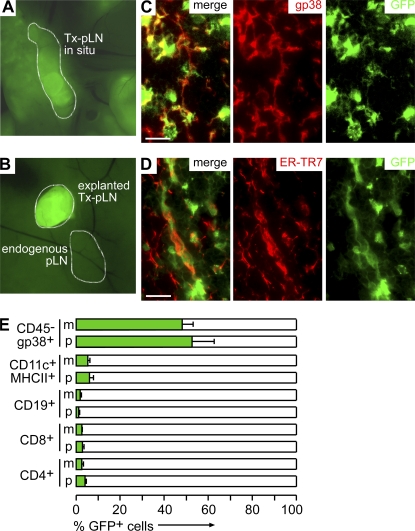
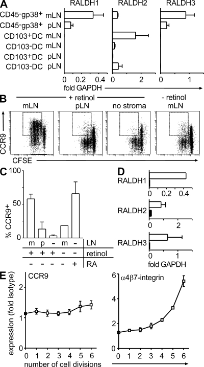
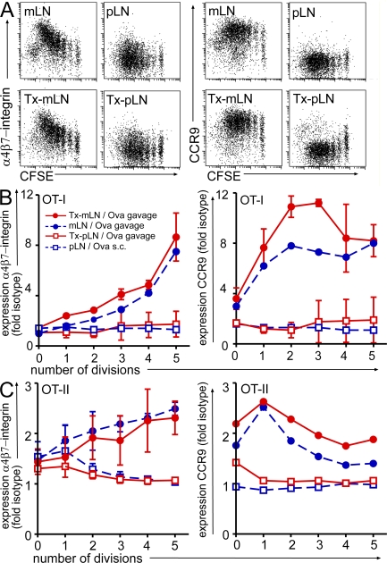
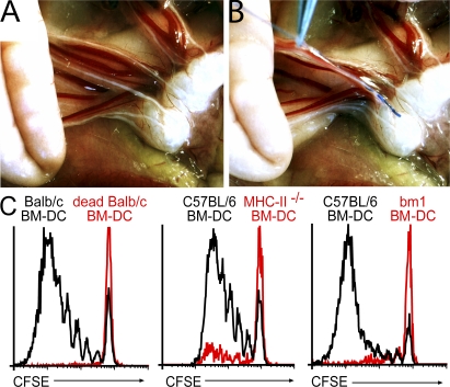
T cells primed in the gut-draining mesenteric lymph nodes (mLN) are imprinted to express alpha4beta7-integrin and chemokine receptor CCR9, thereby enabling lymphocytes to migrate to the small intestine. In vitro activation by intestinal dendritic cells (DC) or addition of retinoic acid (RA) is sufficient to instruct expression of these gut-homing molecules. We report that in vivo stroma cells, but not DC, allow the mLN to induce the generation of gut tropism. Peripheral LN (pLN) transplanted into the gut mesenteries fail to support the generation of gut-homing T cells, even though gut-derived DC enter the transplants and prime T cells. DC that fail to induce alpha4beta7-integrin and CCR9 in vitro readily induce these factors in vivo upon injection into mLN afferent lymphatics. Moreover, uniquely mesenteric but not pLN stroma cells express high levels of RA-producing enzymes and support induction of CCR9 on activated T cells in vitro. These results demonstrate a hitherto unrecognized contribution of stromal cell delivered signals, including RA, on the imprinting of tissue tropism in vivo.
在引流肠道的肠系膜淋巴结(mLN)中被激活的T细胞会被印记上表达α4β7整合素和趋化因子受体CCR9,从而使淋巴细胞能够迁移至小肠。肠道树突状细胞(DC)在体外的激活作用或视黄酸(RA)的添加足以指导这些归巢至肠道的分子的表达。我们报告称,在体内,基质细胞而非DC,使得mLN能够诱导肠道趋向性的产生。移植至肠道系膜中的外周淋巴结(pLN)无法支持归巢至肠道的T细胞的产生,即便源自肠道的DC进入了移植组织并激活了T细胞。在体外无法诱导α4β7整合素和CCR9的DC,在注入mLN输入淋巴管后,能在体内轻易诱导这些因子的产生。此外,唯有肠系膜而非pLN的基质细胞表达高水平的产生RA的酶,并在体外支持激活的T细胞上CCR9的诱导。这些结果证明了基质细胞传递的信号(包括RA)在体内对组织趋向性印记的贡献,而这一贡献此前未被认识到。