Aizenstein Howard J, Butters Meryl A, Wu Minjie, Mazurkewicz Laura M, Stenger V Andrew, Gianaros Peter J, Becker James T, Reynolds Charles F, Carter Cameron S
Department of Psychiatry, University of Pittsburgh, and Western Psychiatric Institute and Clinic, 3811 O'Hara Street, Pittsburgh, PA 15213, USA.
Am J Geriatr Psychiatry. 2009 Jan;17(1):30-42. doi: 10.1097/JGP.0b013e31817b60af.
To characterize the functional neuroanatomy of late-life depression (LLD) by probing for both episodic and persistent alterations in the executive-control circuit of elderly adults.
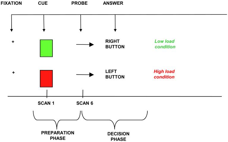
Event-related functional magnetic resonance imaging (fMRI) data were collected while participants performed an executive-control task.
Participants were recruited through a depression-treatment study within the Pittsburgh, PA, Intervention Research Center for Late-Life Mood Disorders.
Thirteen nondepressed elderly comparison participants and 13 LLD patients.
The depressed patients underwent imaging before initiating and after completing 12 weeks of paroxetine.
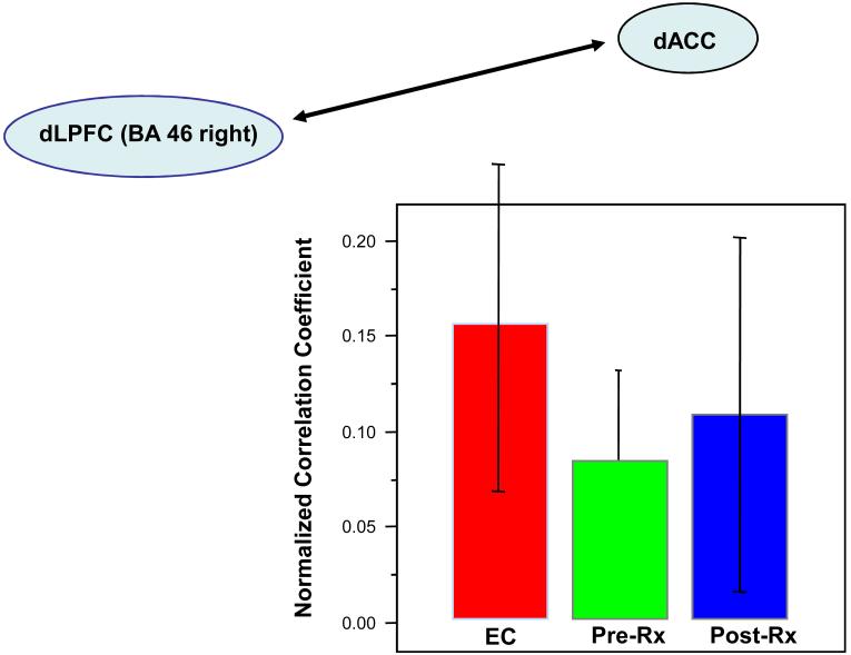
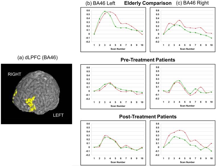
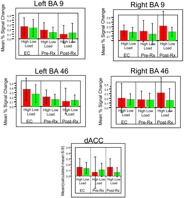
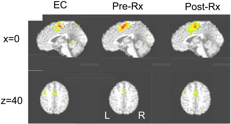
Regional fMRI activity was assessed in the dorsolateral prefrontal cortex (dLPFC: BA9 and BA46 bilaterally) and the dorsal anterior cingulate cortex (dACC). Functional connectivity was assessed by correlating the fMRI time-series in the dLPFC and dACC.
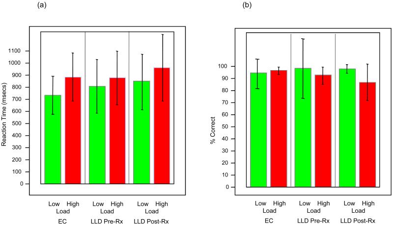
Both depressed and comparison participants performed the task as expected, with greater response latency during high versus low-load trials. The response-latency load-effect did not differ between groups. In contrast to the null findings for behavioral data, pretreatment, depressed patients showed diminished activity in the dLPFC (BA46 left, t(25)=1.9, p = 0.035) and diminished functional connectivity between the dLPFC and dACC. Moreover, right dLPFC (BA46 right, t(25)=2.17, p < 0.02) showed increased activity after treatment.
These results support a model of both episodic and persistent neurobiologic components of LLD. The altered functional connectivity,perhaps due to vascular damage to frontal white matter, appears to be persistent. Further, at least some of the prefrontal hypoactivity (in the right dLPFC) seems to be an episodic characteristic of acute depression amenable to treatment.
通过探究老年人执行控制回路中的发作性和持续性改变,来描述老年期抑郁症(LLD)的功能性神经解剖学特征。
在参与者执行执行控制任务时收集事件相关功能磁共振成像(fMRI)数据。
参与者通过宾夕法尼亚州匹兹堡市晚期生活情绪障碍干预研究中心的一项抑郁症治疗研究招募。
13名无抑郁的老年对照参与者和13名LLD患者。
抑郁症患者在开始服用帕罗西汀前和完成12周治疗后进行成像。
评估背外侧前额叶皮质(双侧BA9和BA46的dLPFC)和背侧前扣带回皮质(dACC)的fMRI区域活动。通过关联dLPFC和dACC中的fMRI时间序列来评估功能连接性。
抑郁组和对照组参与者均按预期完成任务,高负荷试验期间的反应潜伏期长于低负荷试验。两组之间的反应潜伏期负荷效应无差异。与行为数据的阴性结果相反,治疗前,抑郁症患者的dLPFC(左侧BA46,t(25)=1.9,p = 0.035)活动减弱,dLPFC和dACC之间的功能连接性降低。此外,右侧dLPFC(右侧BA46,t(25)=2.17,p < 0.02)在治疗后活动增加。
这些结果支持了LLD发作性和持续性神经生物学成分的模型。功能连接性改变可能是由于额叶白质的血管损伤,似乎是持续性的。此外,至少部分前额叶低活性(右侧dLPFC)似乎是急性抑郁症可治疗的发作性特征。