Soares Janir Alves, Leonardo Mário Roberto, da Silva Léa Assed Bezerra, Tanomaru Filho Mário, Ito Izabel Yoko
Discipline of Endodontics, Department of Dentistry, Federal University of Valleys of Jequitinhonha and Mucuri, Diamantina, MG, Brazil.
J Appl Oral Sci. 2006 Oct;14(5):355-64. doi: 10.1590/s1678-77572006000500011.
The purpose of this study was to evaluate the distribution of microorganisms in the root canal system (RCS) and periapical lesions of dogs' teeth after rotary instrumentation and placement of different calcium hydroxide [Ca(OH)2]-based intracanal dressings.
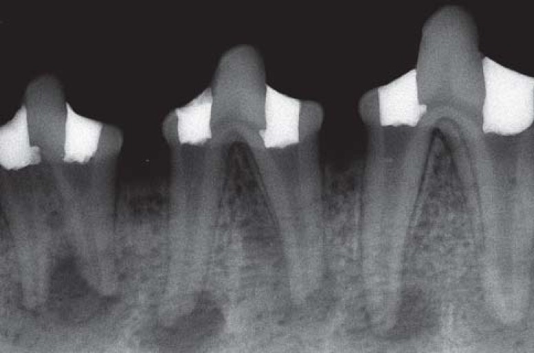
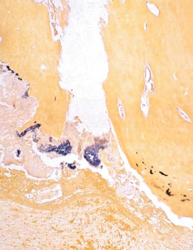
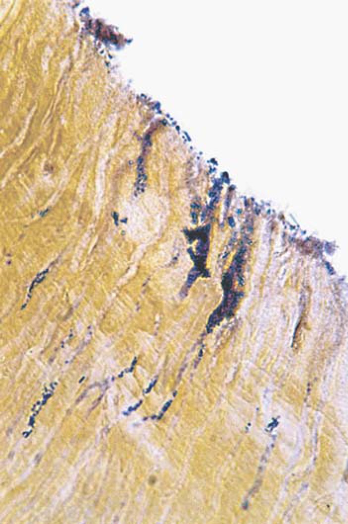
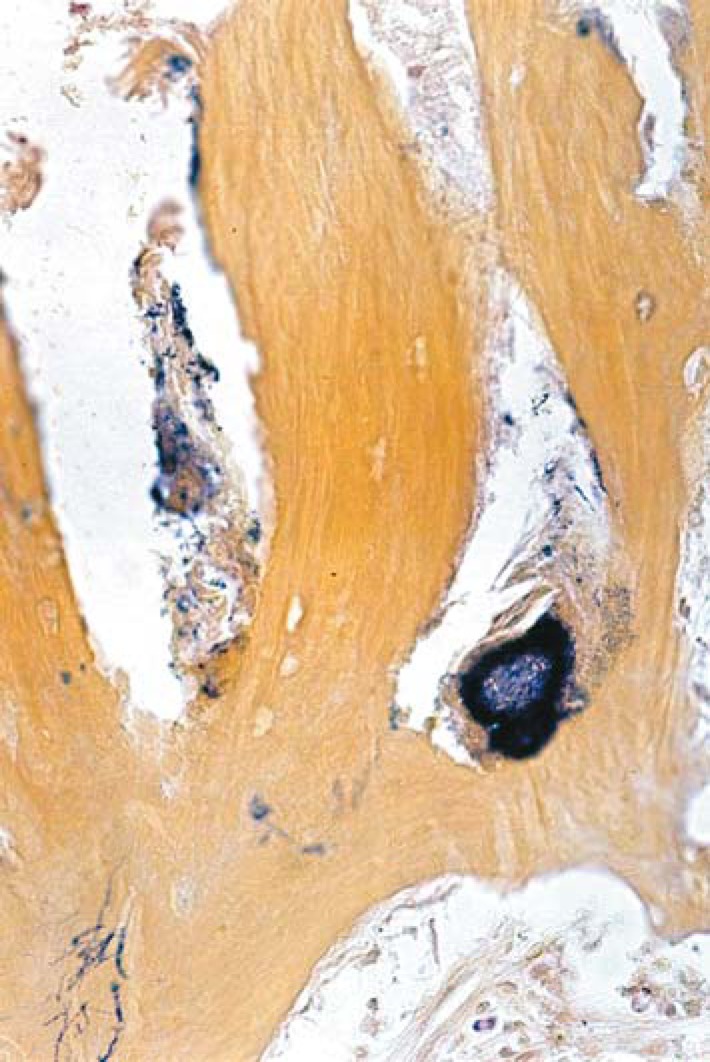
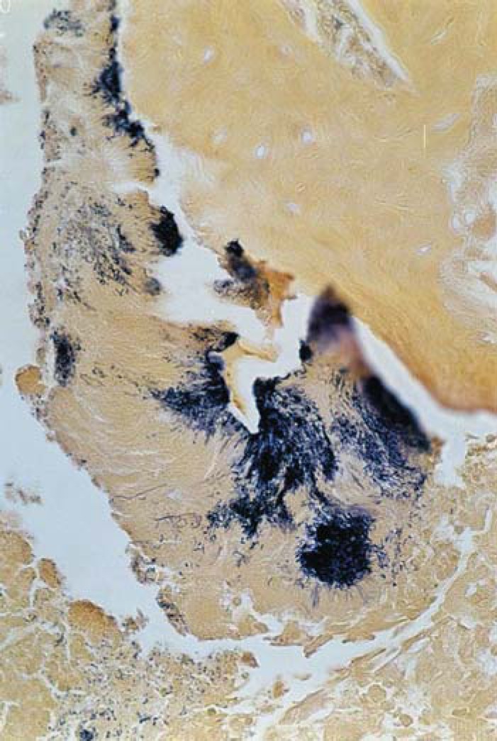
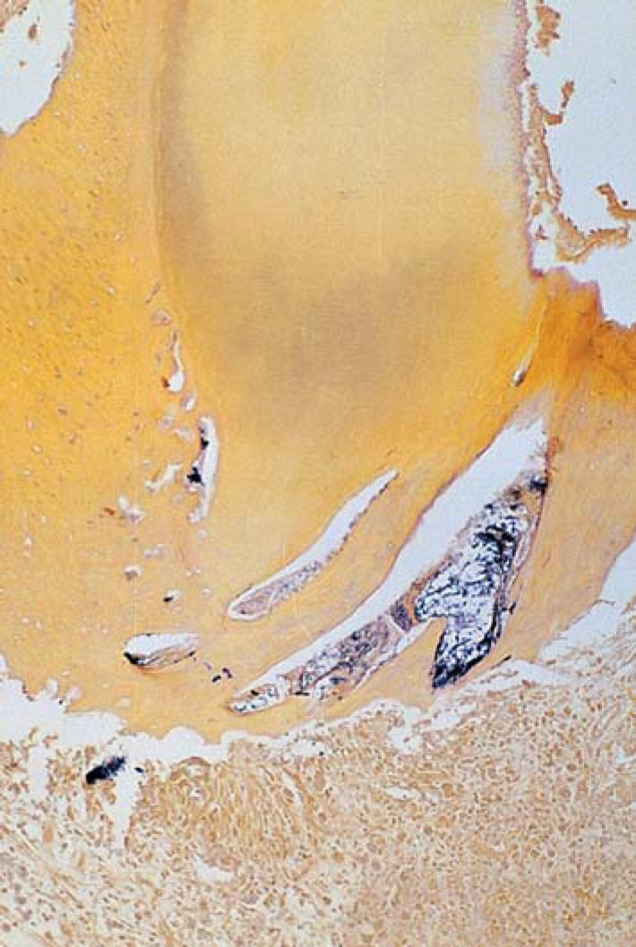
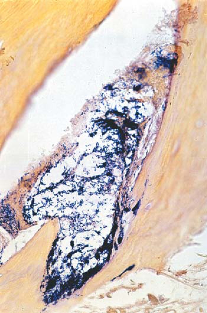
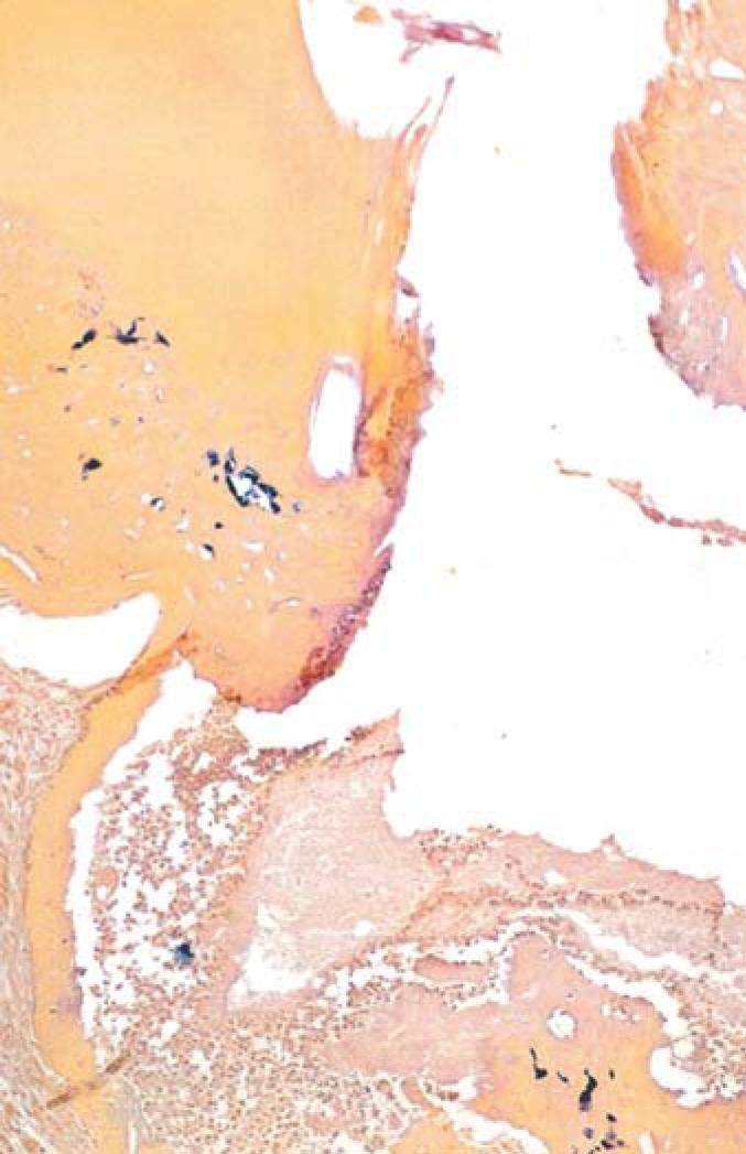
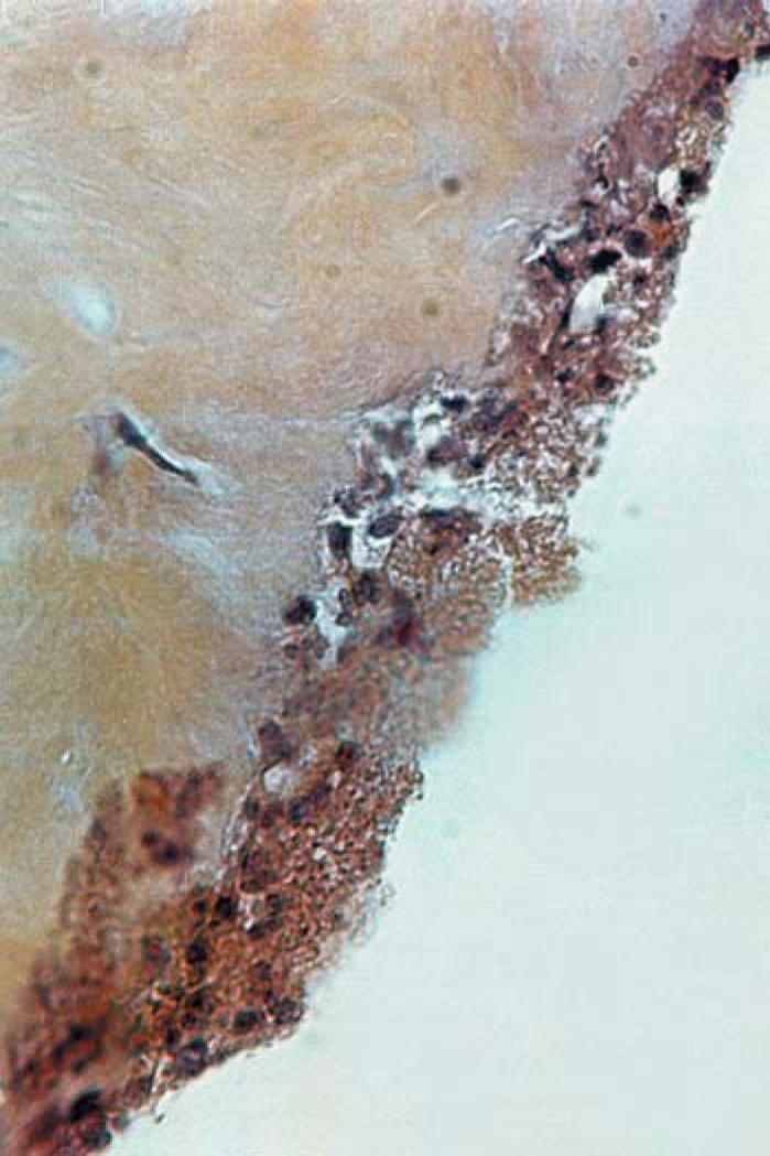
Chronic periapical lesions were experimentally induced in 80 premolar roots of four dogs. Instrumentation was undertaken using the ProFile rotary system and irrigation with 5.25% sodium hypochlorite. The following Ca(OH)2-based pastes were applied for 21 days: group 1 - Calen (n=18); group 2 - Calen+CPMC (n=20); group 3 - Ca(OH)2 p.a. + anaesthetic solution (n=16) and group 4 - Ca(OH)2 p.a.+ 2% chlorhexidine digluconate (n=18). Eight root canals without endodontic treatment constituted the control group. Histological sections were obtained and stained with Brown & Brenn staining technique to evaluate the presence of microorganisms in the main root canal, ramifications of the apical delta and secondary canals, apical cementoplasts, dentinal tubules, areas of cemental resorption and periapical lesions. The results were analyzed statistically by the Mann-Whitney U test (p<0.05).
The control group showed the highest prevalence of microorganisms in all sites evaluated. Gram-positive cocci, bacilli and filaments were the most frequent morphotypes. Similar microbial distribution patterns in the RCS and areas of cementum resorption were observed in all groups (p>0.05). The percentage of RCS sites containing microorganisms in groups 1, 2, 3, 4 and control were: 67.6%, 62.5%, 78.2%, 62.0% and 87.6%, respectively.
In conclusion, the histomicrobiological analysis showed that the rotary instrumentation and the different calcium hydroxide pastes employed did not effectively eliminate the infection from the RCS and periapical lesions. However, several bacteria seen in the histological sections were probably dead or were inactivated by the biomechanical preparation and calcium hydroxide-based intracanal dressing.
本研究旨在评估旋转器械预备及放置不同氢氧化钙[Ca(OH)₂]类根管内封药后犬牙根管系统(RCS)及根尖周病变中微生物的分布情况。
在4只犬的80颗前磨牙牙根上实验性诱导慢性根尖周病变。使用Profile旋转系统进行根管预备,并用5.25%次氯酸钠冲洗。将以下氢氧化钙类糊剂应用21天:第1组 - Calen(n = 18);第2组 - Calen + CPMC(n = 20);第3组 - 氢氧化钙(p.a.)+麻醉剂溶液(n = 16);第4组 - 氢氧化钙(p.a.)+ 2%葡萄糖酸洗必泰(n = 18)。8个未经牙髓治疗的根管作为对照组。获取组织学切片,采用Brown & Brenn染色技术染色,以评估主根管、根尖分歧和侧支根管、根尖牙骨质膜、牙本质小管、牙骨质吸收区域及根尖周病变中微生物的存在情况。结果采用Mann-Whitney U检验进行统计学分析(p < 0.05)。
对照组在所有评估部位的微生物检出率最高。革兰氏阳性球菌、杆菌和丝状菌是最常见的形态类型。所有组在RCS及牙骨质吸收区域均观察到相似的微生物分布模式(p > 0.05)。第1、2、3、4组和对照组中含有微生物的RCS部位百分比分别为:67.6%、62.5%、78.2%、62.0%和87.6%。
总之,组织微生物学分析表明,旋转器械预备及使用的不同氢氧化钙糊剂未能有效消除RCS及根尖周病变中的感染。然而,组织学切片中观察到的几种细菌可能已死亡或因生物力学预备及氢氧化钙类根管内封药而失活。