• 文献检索
  • 文档翻译
  • 深度研究
  • 学术资讯
  • Suppr Zotero 插件Zotero 插件
  • 邀请有礼
  • 套餐&价格
  • 历史记录
应用&插件
Suppr Zotero 插件Zotero 插件浏览器插件Mac 客户端Windows 客户端微信小程序
定价
高级版会员购买积分包购买API积分包
服务
文献检索文档翻译深度研究API 文档MCP 服务
关于我们
关于 Suppr公司介绍联系我们用户协议隐私条款
关注我们

Suppr 超能文献

核心技术专利:CN118964589B侵权必究
粤ICP备2023148730 号-1Suppr @ 2026

文献检索

告别复杂PubMed语法,用中文像聊天一样搜索,搜遍4000万医学文献。AI智能推荐,让科研检索更轻松。

立即免费搜索

文件翻译

保留排版,准确专业,支持PDF/Word/PPT等文件格式,支持 12+语言互译。

免费翻译文档

深度研究

AI帮你快速写综述,25分钟生成高质量综述,智能提取关键信息,辅助科研写作。

立即免费体验

颌骨根尖周病变:伊巴丹104例病例回顾

Periapical lesions of the jaws: a review of 104 cases in ibadan.

作者信息

Akinyamoju A O, Gbadebo S O, Adeyemi B F

机构信息

Department of Oral Pathology, Faculty of Dentistry, College of Medicine, University of Ibadan.

Department of Restorative Dentistry, Faculty of Dentistry, College of Medicine, University of Ibadan.

出版信息

Ann Ib Postgrad Med. 2014 Dec;12(2):115-9.

PMID:25960702
原文链接:https://pmc.ncbi.nlm.nih.gov/articles/PMC4415388/
Abstract

BACKGROUND

Periapical lesions (PLs) occur as a result of pulpal inflammation and may rarely be seen in the absence of pulpal diseases. They are the most common pathological lesions affecting the alveolar bone.

OBJECTIVE

This study aims to describe the clinicopathological features of PLs of the jaws with emphasis on the two most common types.

METHODS

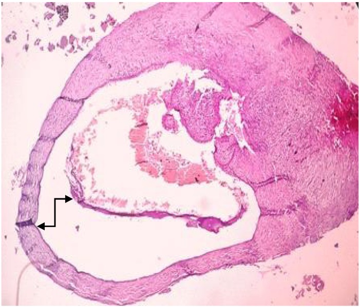
Histopathology records of PLs diagnosed from January 1990 to December 2012 at the Department of Oral Pathology, University College Hospital Ibadan, were examined and categorized into periapical cysts (PCs); periapical granuloma (PGs) and others. Clinical data and histopathological features of these PLs were reviewed and analyzed.

RESULTS

One hundred and four lesions met the criteria for this study and consisted of PGs with 71 (68.3%) cases and PCs with 31 (29.8%) cases and one case each of apical scar and pleomorphic adenoma. Age range of cases was 9 to 80 years (mean=35.6 ± 15.8years) with a peak at age group of 20-29 years. Females were more frequently affected with 51.9% of cases. PLs were most frequently diagnosed in the anterior maxillary region with 58 (56.9%) cases, while the most frequently involved tooth was the left maxillary central incisor with 23 (22.1%) cases.

CONCLUSION

Findings in this study are consistent with those of previous studies. It is important for all periapical pathological specimens to be submitted for histological examination to establish an accurate diagnosis and aid in the identification of sinister lesions that may present in the Periradicular region of teeth.

摘要

背景

根尖周病变(PLs)是牙髓炎症的结果,在无牙髓疾病时很少见。它们是影响牙槽骨的最常见病理病变。

目的

本研究旨在描述颌骨PLs的临床病理特征,重点关注两种最常见的类型。

方法

检查了1990年1月至2012年12月在伊巴丹大学学院医院口腔病理科诊断的PLs的组织病理学记录,并将其分类为根尖周囊肿(PCs)、根尖周肉芽肿(PGs)和其他类型。回顾并分析了这些PLs的临床资料和组织病理学特征。

结果

104个病变符合本研究标准,其中PGs 71例(68.3%),PCs 31例(29.8%),根尖瘢痕和多形性腺瘤各1例。病例年龄范围为9至80岁(平均=35.6±15.8岁),高峰年龄组为20 - 29岁。女性受影响更频繁,占病例的51.9%。PLs最常在前上颌区域诊断出,共58例(56.9%),而最常受累的牙齿是左上颌中切牙,共23例(22.1%)。

结论

本研究结果与先前研究一致。所有根尖周病理标本都应提交进行组织学检查,以建立准确的诊断,并有助于识别可能出现在牙根尖周区域的恶性病变,这一点很重要。

https://cdn.ncbi.nlm.nih.gov/pmc/blobs/8216/4415388/b39f825dede4/AIPM-12-115_F3.jpg
https://cdn.ncbi.nlm.nih.gov/pmc/blobs/8216/4415388/3398db58356a/AIPM-12-115_F1.jpg
https://cdn.ncbi.nlm.nih.gov/pmc/blobs/8216/4415388/b8ee94c1fc9c/AIPM-12-115_F2.jpg
https://cdn.ncbi.nlm.nih.gov/pmc/blobs/8216/4415388/b39f825dede4/AIPM-12-115_F3.jpg
https://cdn.ncbi.nlm.nih.gov/pmc/blobs/8216/4415388/3398db58356a/AIPM-12-115_F1.jpg
https://cdn.ncbi.nlm.nih.gov/pmc/blobs/8216/4415388/b8ee94c1fc9c/AIPM-12-115_F2.jpg
https://cdn.ncbi.nlm.nih.gov/pmc/blobs/8216/4415388/b39f825dede4/AIPM-12-115_F3.jpg

相似文献

1
Periapical lesions of the jaws: a review of 104 cases in ibadan.颌骨根尖周病变:伊巴丹104例病例回顾
Ann Ib Postgrad Med. 2014 Dec;12(2):115-9.
2
Clinicopathological study of 252 jaw bone periapical lesions from a private pathology laboratory.252 例私营病理实验室颌骨根尖周病变的临床病理研究。
J Formos Med Assoc. 2010 Nov;109(11):810-8. doi: 10.1016/S0929-6646(10)60126-X.
3
Periapical lesions are not always a sequelae of pulpal necrosis: a retrospective study of 1521 biopsies.根尖病变并不总是牙髓坏死的后遗症:1521 例活检的回顾性研究。
Int Endod J. 2015 Jan;48(1):68-73. doi: 10.1111/iej.12276. Epub 2014 Apr 2.
4
Clinicopathological comparison of periapical cyst and periapical granuloma in a cohort of Tamil population.泰米尔人群队列中根尖囊肿与根尖肉芽肿的临床病理比较
J Conserv Dent Endod. 2024 May;27(5):524-528. doi: 10.4103/JCDE.JCDE_160_24. Epub 2024 May 10.
5
PERIAPICAL PATHOLOGY: COMPARISON OF CLINICAL DIAGNOSIS AND HISTOPATHOLOGICAL FINDINGS.根尖周病理学:临床诊断与组织病理学发现的比较
J West Afr Coll Surg. 2014 Jul-Sep;4(3):74-88.
6
Melanin pigmented periradicular lesions of extracted teeth: a report of 3 cases and review of the literature.
Afr J Med Med Sci. 2011 Sep;40(3):277-81.
7
The root of the problem: Occurrence of typical and atypical periapical pathoses.问题的根源:典型和非典型根尖周病变的发生。
J Am Dent Assoc. 2016 Aug;147(8):646-9. doi: 10.1016/j.adaj.2016.02.018. Epub 2016 Apr 1.
8
Radiographic and histological evaluation of persistent periapical lesions associated with endodontic failures after apical microsurgery.根尖手术治疗后根管治疗失败相关持续性根尖周病变的放射学和组织学评估。
Int Endod J. 2016 Nov;49(11):1011-1019. doi: 10.1111/iej.12554. Epub 2015 Oct 7.
9
Periapical central giant cell granuloma misdiagnosed as odontogenic cyst.被误诊为牙源性囊肿的根尖中央巨细胞肉芽肿
Int Endod J. 2006 Jun;39(6):510-5. doi: 10.1111/j.1365-2591.2006.01107.x.
10
A Retrospective Brazilian Multicenter Study of Biopsies at the Periapical Area: Identification of Cases of Nonendodontic Periapical Lesions.巴西一项回顾性多中心根尖区活检研究:非牙髓性根尖周病变病例的鉴定。
J Endod. 2020 Apr;46(4):490-495. doi: 10.1016/j.joen.2020.01.003. Epub 2020 Feb 13.

引用本文的文献

1
Is there a higher prevalence of apical periodontitis in head and neck cancer patients? A retrospective study with panoramic radiographs.头颈癌患者根尖周炎的患病率是否更高?一项基于全景X线片的回顾性研究。
Natl J Maxillofac Surg. 2025 Jan-Apr;16(1):39-43. doi: 10.4103/njms.njms_118_24. Epub 2025 Apr 28.
2
Histological and immunohistochemical analysis of human periapical lesions: a study of TGF-β1 and CD68 markers.人根尖周病变的组织学和免疫组织化学分析:转化生长因子-β1和CD68标志物的研究
BMC Oral Health. 2025 Apr 11;25(1):526. doi: 10.1186/s12903-025-05845-2.
3
The Limitations of Periapical X-ray Assessment in Endodontic Diagnosis-A Systematic Review.

本文引用的文献

1
A Twenty-year Survey of Pathologic Reports of Two Common Types of Chronic Periapical Lesions in Shiraz Dental School.设拉子牙科学院两种常见类型慢性根尖周病变病理报告的二十年调查
J Dent Res Dent Clin Dent Prospects. 2008 Spring;2(2):63-70. doi: 10.5681/joddd.2008.013. Epub 2008 Aug 15.
2
Periapical granuloma associated with extracted teeth.与拔除牙齿相关的根尖周肉芽肿。
Niger J Clin Pract. 2011 Jul-Sep;14(3):293-6. doi: 10.4103/1119-3077.86770.
3
Clinical, radiographic and histological evaluation of chronic periapical inflammatory lesions.
根尖X线片评估在牙髓病诊断中的局限性——一项系统评价
J Clin Med. 2023 Jul 12;12(14):4647. doi: 10.3390/jcm12144647.
4
Plasma Rich in Growth Factors in the Treatment of Endodontic Periapical Lesions in Adult Patients: 3-Dimensional Analysis Using Cone-Beam Computed Tomography on the Outcomes of Non-Surgical Endodontic Treatment Using A-PRF+ and Calcium Hydroxide: A Retrospective Cohort Study.富含生长因子血浆治疗成年患者牙髓根尖周病变:使用锥形束计算机断层扫描对使用A-PRF+和氢氧化钙的非手术牙髓治疗结果进行三维分析:一项回顾性队列研究
J Clin Med. 2022 Oct 16;11(20):6092. doi: 10.3390/jcm11206092.
5
PERIAPICAL PATHOLOGY: COMPARISON OF CLINICAL DIAGNOSIS AND HISTOPATHOLOGICAL FINDINGS.根尖周病理学:临床诊断与组织病理学发现的比较
J West Afr Coll Surg. 2014 Jul-Sep;4(3):74-88.
慢性根尖周炎性病变的临床、放射学和组织学评估。
J Appl Oral Sci. 2004 Jun;12(2):117-20. doi: 10.1590/s1678-77572004000200007.
4
Clinicopathological study of 252 jaw bone periapical lesions from a private pathology laboratory.252 例私营病理实验室颌骨根尖周病变的临床病理研究。
J Formos Med Assoc. 2010 Nov;109(11):810-8. doi: 10.1016/S0929-6646(10)60126-X.
5
Histology of periapical lesions obtained during apical surgery.根尖手术中获取的根尖周病变组织学
J Endod. 2009 May;35(5):634-42. doi: 10.1016/j.joen.2009.01.024.
6
Histomicrobiologic aspects of the root canal system and periapical lesions in dogs' teeth after rotary instrumentation and intracanal dressing with Ca(OH)2 pastes.犬牙经旋转器械预备及使用氢氧化钙糊剂进行根管内封药后根管系统及根尖周病变的组织微生物学特征
J Appl Oral Sci. 2006 Oct;14(5):355-64. doi: 10.1590/s1678-77572006000500011.
7
Clinicopathology of soft tissue lesions associated with extracted teeth.与拔牙相关的软组织病变的临床病理学
J Oral Maxillofac Surg. 2008 Nov;66(11):2284-9. doi: 10.1016/j.joms.2008.03.005.
8
The post-endodontic periapical lesion: histologic and etiopathogenic aspects.根管治疗后的根尖周病变:组织学和病因学方面
Med Oral Patol Oral Cir Bucal. 2007 Dec 1;12(8):E585-90.
9
Proliferation of epithelial cell rests, formation of apical cysts, and regression of apical cysts after periapical wound healing.根尖伤口愈合后上皮细胞残余的增殖、根尖囊肿的形成及根尖囊肿的消退。
J Endod. 2007 Aug;33(8):908-16. doi: 10.1016/j.joen.2007.02.006. Epub 2007 Apr 2.
10
Differential diagnosis of large periapical lesions using cone-beam computed tomography measurements and biopsy.使用锥形束计算机断层扫描测量和活检对大根尖周病变进行鉴别诊断。
J Endod. 2006 Sep;32(9):833-7. doi: 10.1016/j.joen.2006.03.008. Epub 2006 Jul 7.