Lee Kuo-Chang, Webb Rick I, Fuerst John A
School of Chemistry and Molecular Biosciences, University of Queensland, Brisbane, Queensland 4072, Australia.
BMC Cell Biol. 2009 Jan 14;10:4. doi: 10.1186/1471-2121-10-4.
Gemmata obscuriglobus is a distinctive member of the divergent phylum Planctomycetes, all known members of which are peptidoglycan-less bacteria with a shared compartmentalized cell structure and divide by a budding process. G. obscuriglobus in addition shares the unique feature that its nucleoid DNA is surrounded by an envelope consisting of two membranes forming an analogous structure to the membrane-bounded nucleoid of eukaryotes and therefore G. obscuriglobus forms a special model for cell biology. Draft genome data for G. obscuriglobus as well as complete genome sequences available so far for other planctomycetes indicate that the key bacterial cell division protein FtsZ is not present in these planctomycetes, so the cell division process in planctomycetes is of special comparative interest. The membrane-bounded nature of the nucleoid in G. obscuriglobus also suggests that special mechanisms for the distribution of this nuclear body to the bud and for distribution of chromosomal DNA might exist during division. It was therefore of interest to examine the cell division cycle in G. obscuriglobus and the process of nucleoid distribution and nuclear body formation during division in this planctomycete bacterium via light and electron microscopy.
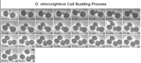
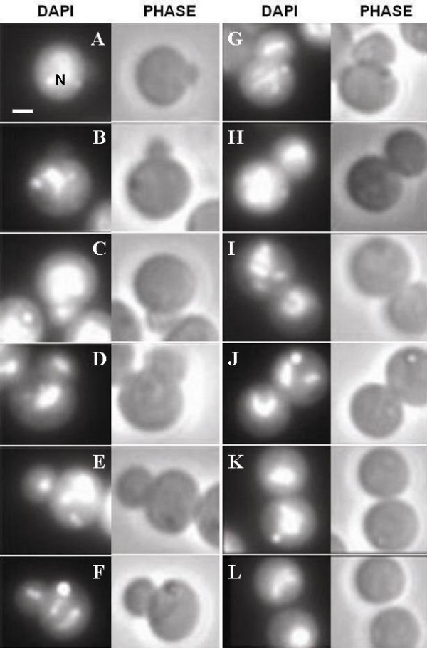
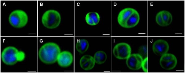
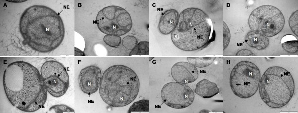
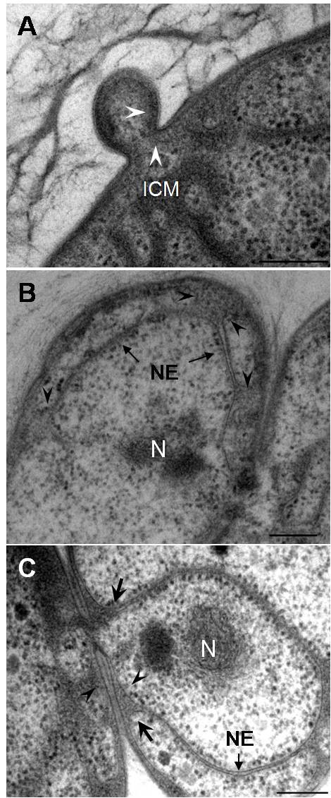
Using phase contrast and fluorescence light microscopy, and transmission electron microscopy, the cell division cycle of G. obscuriglobus was determined. During the budding process, the bud was formed and developed in size from one point of the mother cell perimeter until separation. The matured daughter cell acted as a new mother cell and started its own budding cycle while the mother cell can itself initiate budding repeatedly. Fluorescence microscopy of DAPI-stained cells of G. obscuriglobus suggested that translocation of the nucleoid and formation of the bud did not occur at the same time. Confocal laser scanning light microscopy applied to cells stained for membranes as well as DNA confirmed the behaviour of the nucleoid and nucleoid envelope during cell division. Electron microscopy of cryosubstituted cells confirmed deductions from light microscopy concerning nucleoid presence in relation to the stage of budding, and showed that the nucleoid was observed to occur in both mother and bud cells only at later budding stages. It further suggested that nucleoid envelope formed only after the nucleoid was translocated into the bud, since envelopes only appeared in more mature buds, while naked nucleoids occurred in smaller buds. Nucleoid envelope appeared to originate from the intracytoplasmic membranes (ICM) of both mother cell and bud. There was always a connecting passage between mother cell and bud during the budding process until separation of the two cells. The division cycle of the nucleated planctomycete G. obscuriglobus appears to be a complex process in which chromosomal DNA is transported to the daughter cell bud after initial formation of the bud, and this can be performed repeatedly by a single mother cell.
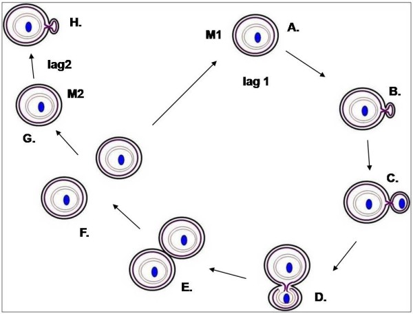
The division cycle of the nucleated planctomycete G. obscuriglobus is a complex process in which chromosomal nucleoid DNA is transported to the daughter cell bud after initial formation of a bud without nucleoid. The new bud nucleoid is initially naked and not surrounded by membrane, but eventually acquires a complete nucleoid envelope consisting of two closely apposed membranes as occurs in the mother cell. The membranes of the new nucleoid envelope surrounding the bud nucleoid are derived from intracytoplasmic membranes of both the mother cell and the bud. The cell division of G. obscuriglobus displays some unique features not known in cells of either prokaryotes or eukaryotes.
暗球菌属(Gemmata obscuriglobus)是进化分支广古菌门(Planctomycetes)中一个独特的成员,该门所有已知成员均为无肽聚糖的细菌,具有共同的区室化细胞结构,并通过出芽过程进行分裂。此外,暗球菌还具有独特的特征,即其拟核DNA被一层由两层膜组成的包膜所包围,形成了与真核生物膜结合拟核类似的结构,因此暗球菌成为细胞生物学的一个特殊模型。暗球菌的基因组草图数据以及目前已有的其他广古菌门细菌的完整基因组序列表明,这些广古菌门细菌中不存在关键的细菌细胞分裂蛋白FtsZ,因此广古菌门细菌的细胞分裂过程具有特殊的比较研究价值。暗球菌中拟核的膜结合性质还表明,在分裂过程中可能存在将这个核体分配到芽中以及分配染色体DNA的特殊机制。因此,通过光学显微镜和电子显微镜研究暗球菌的细胞分裂周期以及该广古菌门细菌在分裂过程中的拟核分布和核体形成过程很有意义。
利用相差显微镜、荧光显微镜和透射电子显微镜确定了暗球菌的细胞分裂周期。在出芽过程中,芽从母细胞周边的一个点开始形成并逐渐长大,直至与母细胞分离。成熟的子细胞作为一个新的母细胞开始其自身的出芽周期,而母细胞本身可以反复启动出芽过程。对暗球菌经4',6-二脒基-2-苯基吲哚(DAPI)染色的细胞进行荧光显微镜观察表明,拟核的易位和芽的形成并非同时发生。应用于膜和DNA染色细胞的共聚焦激光扫描显微镜证实了细胞分裂过程中拟核和拟核包膜的行为。对冷冻置换细胞的电子显微镜观察证实了光学显微镜关于拟核在出芽阶段存在情况的推断,并表明仅在出芽后期才能在母细胞和芽细胞中同时观察到拟核。这进一步表明,拟核包膜仅在拟核易位到芽中之后形成,因为包膜仅出现在更成熟的芽中,而较小的芽中则存在裸露的拟核。拟核包膜似乎起源于母细胞和芽的胞内膜(ICM)。在出芽过程中,直到两个细胞分离之前,母细胞和芽之间始终存在连接通道。有核广古菌门细菌暗球菌的分裂周期似乎是一个复杂的过程,其中染色体DNA在芽最初形成后被转运到子细胞芽中,并且单个母细胞可以反复进行这一过程。
有核广古菌门细菌暗球菌的分裂周期是一个复杂的过程,其中染色体拟核DNA在无拟核的芽最初形成后被转运到子细胞芽中。新的芽拟核最初是裸露的,没有被膜包围,但最终会获得一个由两层紧密贴合的膜组成的完整拟核包膜,就像母细胞中的情况一样。围绕芽拟核的新拟核包膜的膜源自母细胞和芽的胞内膜。暗球菌的细胞分裂表现出一些原核生物或真核生物细胞中所没有的独特特征。