Sirotin Yevgeniy B, Das Aniruddha
Department of Neuroscience, Columbia University, New York, New York 10027, USA.
Nature. 2009 Jan 22;457(7228):475-9. doi: 10.1038/nature07664.
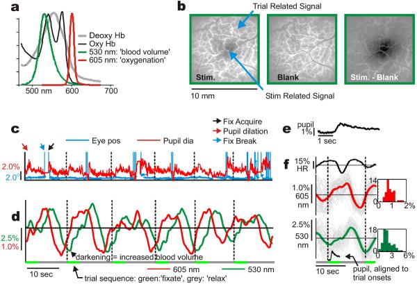
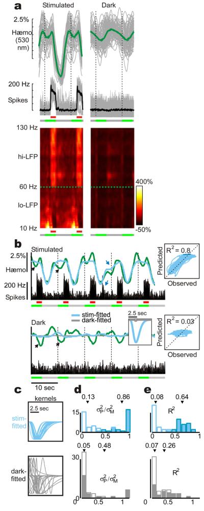
Haemodynamic signals underlying functional brain imaging (for example, functional magnetic resonance imaging (fMRI)) are assumed to reflect metabolic demand generated by local neuronal activity, with equal increases in haemodynamic signal implying equal increases in the underlying neuronal activity. Few studies have compared neuronal and haemodynamic signals in alert animals to test for this assumed correspondence. Here we present evidence that brings this assumption into question. Using a dual-wavelength optical imaging technique that independently measures cerebral blood volume and oxygenation, continuously, in alert behaving monkeys, we find two distinct components to the haemodynamic signal in the alert animals' primary visual cortex (V1). One component is reliably predictable from neuronal responses generated by visual input. The other component-of almost comparable strength-is a hitherto unknown signal that entrains to task structure independently of visual input or of standard neural predictors of haemodynamics. This latter component shows predictive timing, with increases of cerebral blood volume in anticipation of trial onsets even in darkness. This trial-locked haemodynamic signal could be due to an accompanying V1 arterial pumping mechanism, closely matched in time, with peaks of arterial dilation entrained to predicted trial onsets. These findings (tested in two animals) challenge the current understanding of the link between brain haemodynamics and local neuronal activity. They also suggest the existence of a novel preparatory mechanism in the brain that brings additional arterial blood to cortex in anticipation of expected tasks.
功能性脑成像(例如,功能磁共振成像(fMRI))所依据的血流动力学信号被认为反映了局部神经元活动产生的代谢需求,血流动力学信号的同等增加意味着潜在神经元活动的同等增加。很少有研究在清醒动物中比较神经元和血流动力学信号以检验这种假设的对应关系。在此,我们提供的证据对这一假设提出了质疑。我们使用一种双波长光学成像技术,在清醒行为的猴子中连续独立测量脑血容量和氧合情况,发现清醒动物初级视觉皮层(V1)的血流动力学信号有两个不同的成分。一个成分可从视觉输入产生的神经元反应可靠地预测出来。另一个强度几乎相当的成分是一个迄今未知的信号,它与任务结构同步,独立于视觉输入或血流动力学的标准神经预测因子。后一个成分显示出预测性的时间关系,即使在黑暗中,在试验开始前脑血容量也会增加。这种与试验锁定的血流动力学信号可能是由于伴随的V1动脉泵血机制,在时间上紧密匹配,动脉扩张峰值与预测的试验开始同步。这些发现(在两只动物身上进行了测试)挑战了目前对脑血流动力学与局部神经元活动之间联系的理解。它们还表明大脑中存在一种新的准备机制,在预期到预期任务时会将额外的动脉血输送到皮层。