Madani Rana, Karastergiou Kalypso, Ogston Nicola C, Miheisi Nazar, Bhome Rahul, Haloob Nora, Tan Garry D, Karpe Fredrik, Malone-Lee James, Hashemi Majid, Jahangiri Marjan, Mohamed-Ali Vidya
Centre for Clinical Pharmacology, Div. of Medicine, University College London, 5 University St., London, UK WC1 6JJ.
Am J Physiol Endocrinol Metab. 2009 Jun;296(6):E1262-8. doi: 10.1152/ajpendo.90511.2008. Epub 2009 Feb 24.
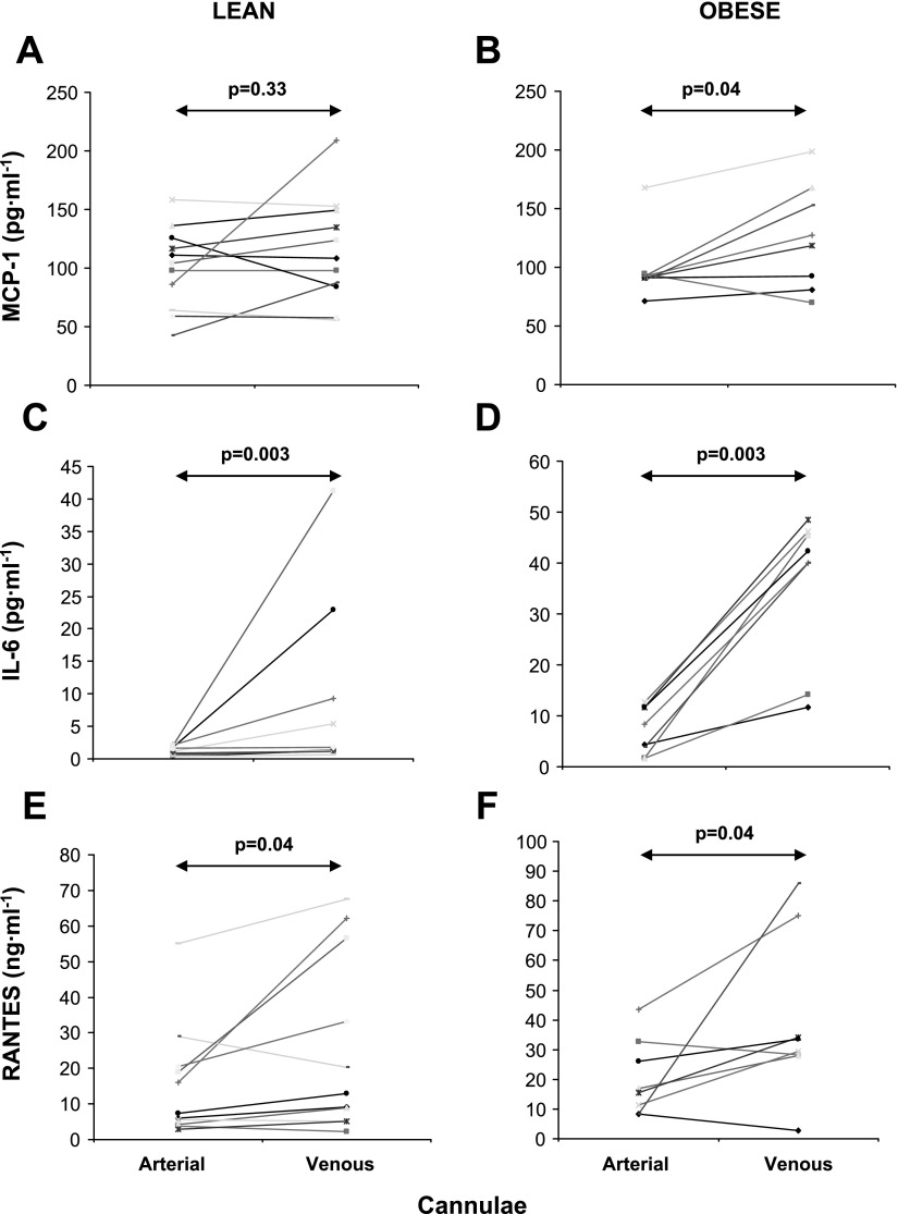
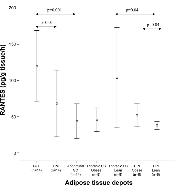
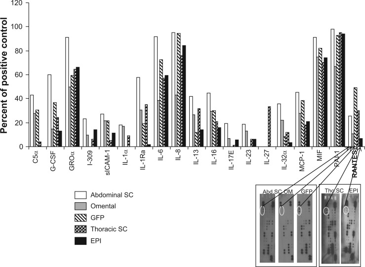
Obesity is associated with elevated inflammatory signals from various adipose tissue depots. This study aimed to evaluate release of regulated on activation, normal T cell expressed and secreted (RANTES) by human adipose tissue in vivo and ex vivo, in reference to monocyte chemoattractant protein-1 (MCP-1) and interleukin-6 (IL-6) release. Arteriovenous differences of RANTES, MCP-1, and IL-6 were studied in vivo across the abdominal subcutaneous adipose tissue in healthy Caucasian subjects with a wide range of adiposity. Systemic levels and ex vivo RANTES release were studied in abdominal subcutaneous, gastric fat pad, and omental adipose tissue from morbidly obese bariatric surgery patients and in thoracic subcutaneous and epicardial adipose tissue from cardiac surgery patients without coronary artery disease. Arteriovenous studies confirmed in vivo RANTES and IL-6 release in adipose tissue of lean and obese subjects and release of MCP-1 in obesity. However, in vivo release of MCP-1 and RANTES, but not IL-6, was lower than circulating levels. Ex vivo release of RANTES was greater from the gastric fat pad compared with omental (P = 0.01) and subcutaneous (P = 0.001) tissue. Epicardial adipose tissue released less RANTES than thoracic subcutaneous adipose tissue in lean (P = 0.04) but not obese subjects. Indexes of obesity correlated with epicardial RANTES but not with systemic RANTES or its release from other depots. In conclusion, RANTES is released by human subcutaneous adipose tissue in vivo and in varying amounts by other depots ex vivo. While it appears unlikely that the adipose organ contributes significantly to circulating levels, local implications of this chemokine deserve further investigation.
肥胖与来自不同脂肪组织储存部位的炎症信号升高有关。本研究旨在参照单核细胞趋化蛋白-1(MCP-1)和白细胞介素-6(IL-6)的释放情况,评估人体脂肪组织在体内和体外释放调节激活正常T细胞表达和分泌因子(RANTES)的情况。在具有广泛肥胖程度的健康白种人受试者中,研究了RANTES、MCP-1和IL-6在腹部皮下脂肪组织中的动静脉差异。对病态肥胖的减肥手术患者的腹部皮下、胃脂肪垫和网膜脂肪组织,以及无冠状动脉疾病的心脏手术患者的胸部皮下和心外膜脂肪组织,研究了全身水平和体外RANTES释放情况。动静脉研究证实,瘦人和肥胖受试者的脂肪组织在体内释放RANTES和IL-6,肥胖者体内释放MCP-1。然而,MCP-1和RANTES的体内释放量低于循环水平,但IL-6并非如此。与网膜组织(P = 0.01)和皮下组织(P = 0.001)相比,胃脂肪垫体外释放的RANTES更多。在心外膜脂肪组织中,瘦人(P = 0.04)释放的RANTES比胸部皮下脂肪组织少,但肥胖受试者并非如此。肥胖指标与心外膜RANTES相关,但与全身RANTES或其从其他储存部位的释放无关。总之,RANTES由人体皮下脂肪组织在体内释放,其他储存部位在体外以不同量释放。虽然脂肪器官似乎不太可能对循环水平有显著贡献,但这种趋化因子的局部影响值得进一步研究。