• 文献检索
  • 文档翻译
  • 深度研究
  • 学术资讯
  • Suppr Zotero 插件Zotero 插件
  • 邀请有礼
  • 套餐&价格
  • 历史记录
应用&插件
Suppr Zotero 插件Zotero 插件浏览器插件Mac 客户端Windows 客户端微信小程序
定价
高级版会员购买积分包购买API积分包
服务
文献检索文档翻译深度研究API 文档MCP 服务
关于我们
关于 Suppr公司介绍联系我们用户协议隐私条款
关注我们

Suppr 超能文献

核心技术专利:CN118964589B侵权必究
粤ICP备2023148730 号-1Suppr @ 2026

文献检索

告别复杂PubMed语法,用中文像聊天一样搜索,搜遍4000万医学文献。AI智能推荐,让科研检索更轻松。

立即免费搜索

文件翻译

保留排版,准确专业,支持PDF/Word/PPT等文件格式,支持 12+语言互译。

免费翻译文档

深度研究

AI帮你快速写综述,25分钟生成高质量综述,智能提取关键信息,辅助科研写作。

立即免费体验

基于监督学习和无监督学习的乳腺MRI可疑病变无模型可视化

Model-Free Visualization of Suspicious Lesions in Breast MRI Based on Supervised and Unsupervised Learning.

作者信息

Twellmann Thorsten, Meyer-Baese Anke, Lange Oliver, Foo Simon, Nattkemper Tim W

机构信息

Department of Electrical and Computer Engineering, Florida State University, Tallahassee, Florida 32310-6046.

出版信息

Eng Appl Artif Intell. 2008 Mar;21(2):129-140. doi: 10.1016/j.engappai.2007.04.005.

DOI:10.1016/j.engappai.2007.04.005
PMID:19255616
原文链接:https://pmc.ncbi.nlm.nih.gov/articles/PMC2597847/
Abstract

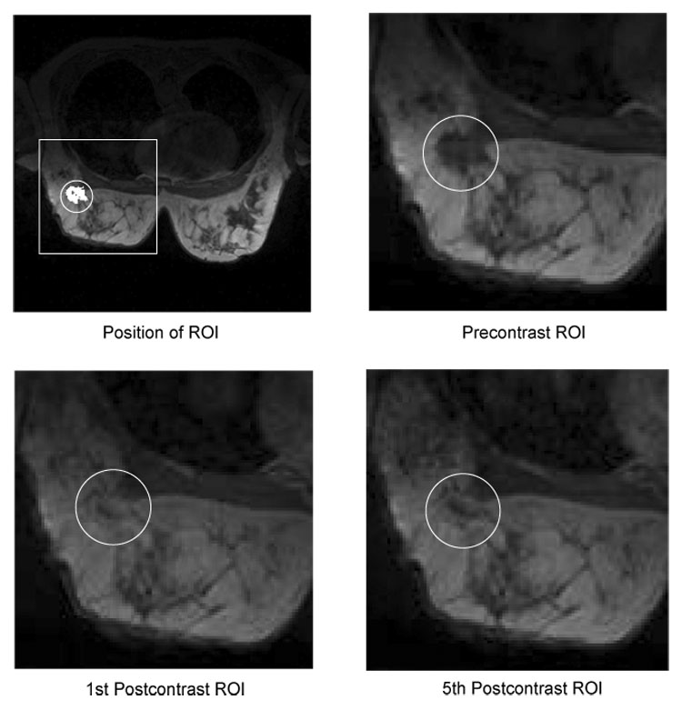
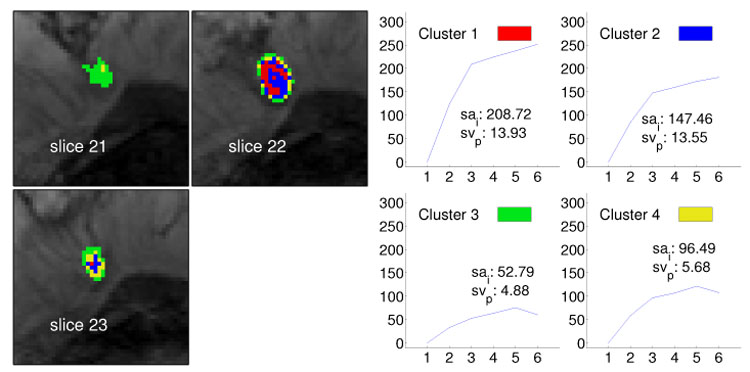
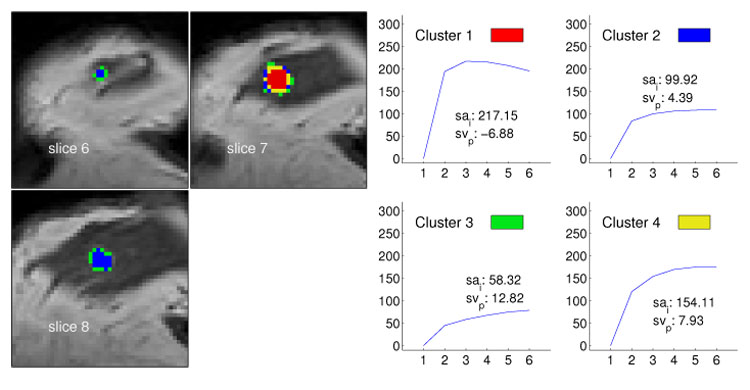
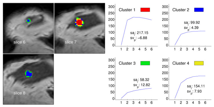
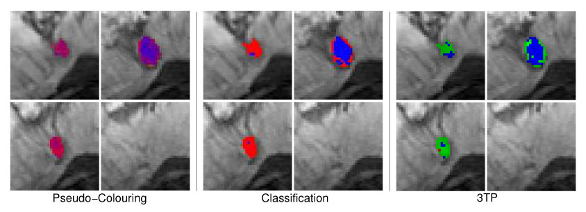
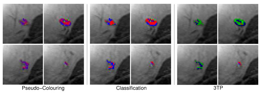
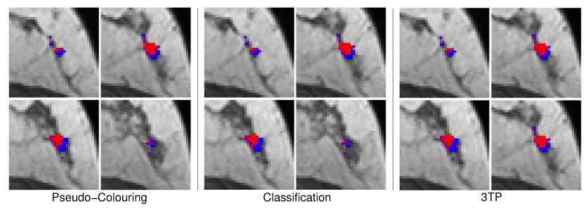
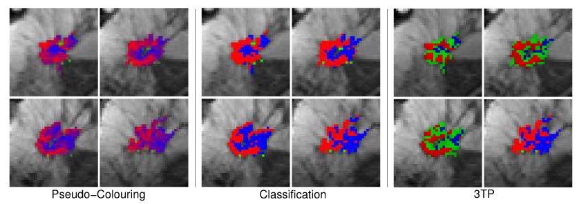
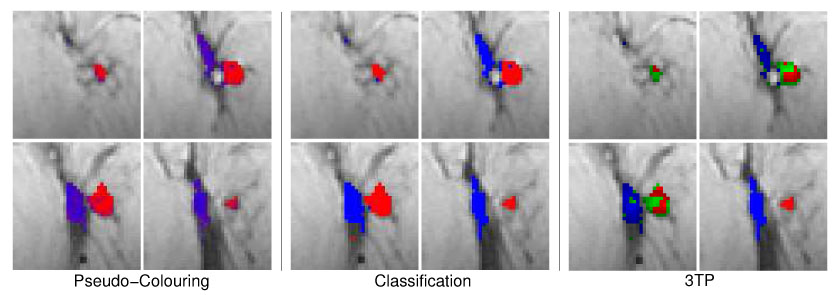
Dynamic contrast-enhanced magnetic resonance imaging (DCE-MRI) has become an important tool in breast cancer diagnosis, but evaluation of multitemporal 3D image data holds new challenges for human observers. To aid the image analysis process, we apply supervised and unsupervised pattern recognition techniques for computing enhanced visualizations of suspicious lesions in breast MRI data. These techniques represent an important component of future sophisticated computer-aided diagnosis (CAD) systems and support the visual exploration of spatial and temporal features of DCE-MRI data stemming from patients with confirmed lesion diagnosis. By taking into account the heterogeneity of cancerous tissue, these techniques reveal signals with malignant, benign and normal kinetics. They also provide a regional subclassification of pathological breast tissue, which is the basis for pseudo-color presentations of the image data. Intelligent medical systems are expected to have substantial implications in healthcare politics by contributing to the diagnosis of indeterminate breast lesions by non-invasive imaging.

摘要

动态对比增强磁共振成像(DCE-MRI)已成为乳腺癌诊断的重要工具,但对多时间点三维图像数据的评估给人类观察者带来了新的挑战。为辅助图像分析过程,我们应用监督和非监督模式识别技术来计算乳腺MRI数据中可疑病变的增强可视化图像。这些技术是未来先进的计算机辅助诊断(CAD)系统的重要组成部分,并支持对确诊病变患者的DCE-MRI数据的空间和时间特征进行可视化探索。通过考虑癌组织的异质性,这些技术可揭示具有恶性、良性和正常动力学的信号。它们还提供了病理性乳腺组织的区域亚分类,这是图像数据伪彩色呈现的基础。智能医疗系统有望通过无创成像对不确定乳腺病变的诊断做出贡献,从而在医疗保健政策方面产生重大影响。

https://cdn.ncbi.nlm.nih.gov/pmc/blobs/61c8/2597847/61210102ced3/nihms45393f12.jpg
https://cdn.ncbi.nlm.nih.gov/pmc/blobs/61c8/2597847/b4e0b8a192f1/nihms45393f1.jpg
https://cdn.ncbi.nlm.nih.gov/pmc/blobs/61c8/2597847/0c5d09f4797c/nihms45393f2.jpg
https://cdn.ncbi.nlm.nih.gov/pmc/blobs/61c8/2597847/c8b2381fa581/nihms45393f3.jpg
https://cdn.ncbi.nlm.nih.gov/pmc/blobs/61c8/2597847/9b18203544a2/nihms45393f4.jpg
https://cdn.ncbi.nlm.nih.gov/pmc/blobs/61c8/2597847/274983a2c198/nihms45393f5.jpg
https://cdn.ncbi.nlm.nih.gov/pmc/blobs/61c8/2597847/1afb5f400472/nihms45393f6.jpg
https://cdn.ncbi.nlm.nih.gov/pmc/blobs/61c8/2597847/6bf69ba26107/nihms45393f7.jpg
https://cdn.ncbi.nlm.nih.gov/pmc/blobs/61c8/2597847/4f732b495201/nihms45393f8.jpg
https://cdn.ncbi.nlm.nih.gov/pmc/blobs/61c8/2597847/c7de9884b51b/nihms45393f9.jpg
https://cdn.ncbi.nlm.nih.gov/pmc/blobs/61c8/2597847/b548a8a31b0e/nihms45393f10.jpg
https://cdn.ncbi.nlm.nih.gov/pmc/blobs/61c8/2597847/52bebb94de65/nihms45393f11.jpg
https://cdn.ncbi.nlm.nih.gov/pmc/blobs/61c8/2597847/61210102ced3/nihms45393f12.jpg

相似文献

1
Model-Free Visualization of Suspicious Lesions in Breast MRI Based on Supervised and Unsupervised Learning.基于监督学习和无监督学习的乳腺MRI可疑病变无模型可视化
Eng Appl Artif Intell. 2008 Mar;21(2):129-140. doi: 10.1016/j.engappai.2007.04.005.
2
An adaptive tissue characterization network for model-free visualization of dynamic contrast-enhanced magnetic resonance image data.一种用于动态对比增强磁共振图像数据无模型可视化的自适应组织表征网络。
IEEE Trans Med Imaging. 2005 Oct;24(10):1256-66. doi: 10.1109/TMI.2005.854517.
3
Potential of computer-aided diagnosis of high spectral and spatial resolution (HiSS) MRI in the classification of breast lesions.高光谱和空间分辨率(HiSS)MRI 计算机辅助诊断在乳腺病变分类中的应用潜力。
J Magn Reson Imaging. 2014 Jan;39(1):59-67. doi: 10.1002/jmri.24145. Epub 2013 Sep 10.
4
A computer-aided diagnosis system for breast DCE-MRI at high spatiotemporal resolution.一种用于高时空分辨率乳腺动态对比增强磁共振成像的计算机辅助诊断系统。
Med Phys. 2016 Jan;43(1):84. doi: 10.1118/1.4937787.
5
Small lesions evaluation based on unsupervised cluster analysis of signal-intensity time courses in dynamic breast MRI.基于动态乳腺磁共振成像中信号强度时间历程的无监督聚类分析对小病变进行评估。
Int J Biomed Imaging. 2009;2009:326924. doi: 10.1155/2009/326924. Epub 2010 Apr 1.
6
A quantitative heterogeneity analysis approach to molecular subtype recognition of breast cancer in dynamic contrast-enhanced magnetic imaging images from radiomics data.一种基于放射组学数据的动态对比增强磁共振成像图像中乳腺癌分子亚型识别的定量异质性分析方法。
Quant Imaging Med Surg. 2023 Jul 1;13(7):4429-4446. doi: 10.21037/qims-22-1230. Epub 2023 May 26.
7
Time-dependent diffusion MRI and kinetic heterogeneity as potential imaging biomarkers for diagnosing suspicious breast lesions with 3.0-T breast MRI.基于3.0-T乳腺MRI的时间依赖性扩散加权成像及动力学异质性作为诊断可疑乳腺病变的潜在影像生物标志物
Magn Reson Imaging. 2025 Apr;117:110323. doi: 10.1016/j.mri.2025.110323. Epub 2025 Jan 4.
8
Multimodality computer-aided breast cancer diagnosis with FFDM and DCE-MRI.基于 FFDM 和 DCE-MRI 的多模态计算机辅助乳腺癌诊断。
Acad Radiol. 2010 Sep;17(9):1158-67. doi: 10.1016/j.acra.2010.04.015.
9
Semi-automatic region-of-interest segmentation based computer-aided diagnosis of mass lesions from dynamic contrast-enhanced magnetic resonance imaging based breast cancer screening.基于动态对比增强磁共振成像的乳腺癌筛查中,基于半自动感兴趣区域分割的计算机辅助乳腺肿块病变诊断。
J Digit Imaging. 2014 Oct;27(5):670-8. doi: 10.1007/s10278-014-9723-y.
10
COMPUTER-AIDED DIAGNOSIS AND VISUALIZATION BASED ON CLUSTERING AND INDEPENDENT COMPONENT ANALYSIS FOR BREAST MRI.基于聚类和独立成分分析的乳腺磁共振成像计算机辅助诊断与可视化
Proc Int Conf Image Proc. 2008 Oct 12;2008:3000-3003. doi: 10.1109/ICIP.2008.4712426.

引用本文的文献

1
Dynamic contrast-enhanced MRI-based radiomics model of intra-tumoral kinetic heterogeneity for predicting breast cancer molecular subtypes.基于动态对比增强磁共振成像的瘤内动力学异质性放射组学模型预测乳腺癌分子亚型
Front Mol Biosci. 2025 Jul 18;12:1635296. doi: 10.3389/fmolb.2025.1635296. eCollection 2025.
2
Appearance Constrained Semi-Automatic Segmentation from DCE-MRI is Reproducible and Feasible for Breast Cancer Radiomics: A Feasibility Study.基于外观约束的 DCE-MRI 半自动分割方法对于乳腺癌放射组学是可重现且可行的:一项可行性研究。
Sci Rep. 2018 Mar 19;8(1):4838. doi: 10.1038/s41598-018-22980-9.
3
Tensor based multichannel reconstruction for breast tumours identification from DCE-MRIs.

本文引用的文献

1
Detection of suspicious lesions in dynamic contrast enhanced MRI data.动态对比增强磁共振成像数据中可疑病变的检测。
Conf Proc IEEE Eng Med Biol Soc. 2004;2006:454-7. doi: 10.1109/IEMBS.2004.1403192.
2
A fuzzy c-means (FCM)-based approach for computerized segmentation of breast lesions in dynamic contrast-enhanced MR images.一种基于模糊C均值(FCM)的方法,用于动态对比增强磁共振图像中乳腺病变的计算机化分割。
Acad Radiol. 2006 Jan;13(1):63-72. doi: 10.1016/j.acra.2005.08.035.
3
An adaptive tissue characterization network for model-free visualization of dynamic contrast-enhanced magnetic resonance image data.
基于张量的多通道重建用于从动态对比增强磁共振成像中识别乳腺肿瘤。
PLoS One. 2017 Mar 10;12(3):e0172111. doi: 10.1371/journal.pone.0172111. eCollection 2017.
4
Pattern identification of biomedical images with time series: Contrasting THz pulse imaging with DCE-MRIs.基于时间序列的生物医学图像模式识别:太赫兹脉冲成像与动态对比增强磁共振成像的对比
Artif Intell Med. 2016 Feb;67:1-23. doi: 10.1016/j.artmed.2016.01.005. Epub 2016 Feb 16.
5
A vector machine formulation with application to the computer-aided diagnosis of breast cancer from DCE-MRI screening examinations.一种应用于基于动态对比增强磁共振成像(DCE-MRI)筛查检查的乳腺癌计算机辅助诊断的向量机公式。
J Digit Imaging. 2014 Feb;27(1):145-51. doi: 10.1007/s10278-013-9621-8.
6
Spectral embedding based active contour (SEAC) for lesion segmentation on breast dynamic contrast enhanced magnetic resonance imaging.基于谱嵌入的主动轮廓(SEAC)用于乳腺动态对比增强磁共振成像的病变分割。
Med Phys. 2013 Mar;40(3):032305. doi: 10.1118/1.4790466.
7
Comparative analysis of nonlinear dimensionality reduction techniques for breast MRI segmentation.比较分析用于乳腺 MRI 分割的非线性降维技术。
Med Phys. 2012 Apr;39(4):2275-89. doi: 10.1118/1.3682173.
8
Textural kinetics: a novel dynamic contrast-enhanced (DCE)-MRI feature for breast lesion classification.纹理动力学:一种新的动态对比增强(DCE)MRI 特征,用于乳腺病变分类。
J Digit Imaging. 2011 Jun;24(3):446-63. doi: 10.1007/s10278-010-9298-1.
一种用于动态对比增强磁共振图像数据无模型可视化的自适应组织表征网络。
IEEE Trans Med Imaging. 2005 Oct;24(10):1256-66. doi: 10.1109/TMI.2005.854517.
4
Dynamic magnetic resonance imaging of tumor perfusion. Approaches and biomedical challenges.肿瘤灌注的动态磁共振成像。方法与生物医学挑战。
IEEE Eng Med Biol Mag. 2004 Sep-Oct;23(5):65-83. doi: 10.1109/memb.2004.1360410.
5
Interactive detection and visualization of breast lesions from dynamic contrast enhanced MRI volumes.基于动态对比增强磁共振成像容积数据的乳腺病变交互式检测与可视化
Comput Med Imaging Graph. 2004 Dec;28(8):435-44. doi: 10.1016/j.compmedimag.2004.07.004.
6
Image fusion for dynamic contrast enhanced magnetic resonance imaging.动态对比增强磁共振成像的图像融合
Biomed Eng Online. 2004 Oct 19;3(1):35. doi: 10.1186/1475-925X-3-35.
7
Independent component analysis for the examination of dynamic contrast-enhanced breast magnetic resonance imaging data: preliminary study.用于检查动态对比增强乳腺磁共振成像数据的独立成分分析:初步研究
Invest Radiol. 2002 Dec;37(12):647-54. doi: 10.1097/00004424-200212000-00002.
8
Clinical testing of high-spatial-resolution parametric contrast-enhanced MR imaging of the breast.
AJR Am J Roentgenol. 2002 Dec;179(6):1485-92. doi: 10.2214/ajr.179.6.1791485.
9
Neural network-based segmentation of dynamic MR mammographic images.
Magn Reson Imaging. 2002 Feb;20(2):147-54. doi: 10.1016/s0730-725x(02)00464-2.
10
Feature extraction and classification of dynamic contrast-enhanced T2*-weighted breast image data.动态对比增强T2*加权乳腺图像数据的特征提取与分类
IEEE Trans Med Imaging. 2001 Dec;20(12):1293-301. doi: 10.1109/42.974924.