Cho Young Jae, Won Jung Bin, Byeon Suk Ho, Yang Woo Ik, Koh Hyoung Jun, Kwon Oh Woong, Lee Sung Chul
Siloam Eye Hospital, and The Institute of Vision Research, Department of Ophthalmology, Yonsei University College of Medicine, Seoul, Korea.
Korean J Ophthalmol. 2009 Mar;23(1):49-52. doi: 10.3341/kjo.2009.23.1.49. Epub 2009 Mar 9.
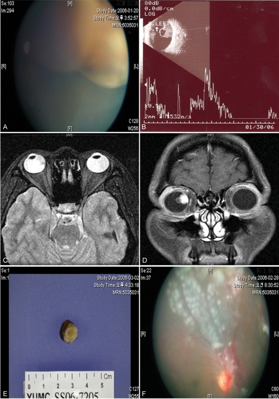
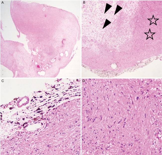
Schwannomas rarely present as intraocular tumors and are often misdiagnosed as malignant melanoma. We describe a choroidal schwannoma confirmed by sclerouvectomy. A 30-year-old woman presented with a large nonpigmented intraocular mass of the choroid in the right eye and underwent surgical excision by sclerouvectomy. Histologically, the tumor was composed of a mixture of cellular solid components (Antoni A) and loose myxoid components (Antoni B). The tumor was eventually diagnosed as a schwannoma. Currently available ancillary studies are still of little value in definitively differentiating schwannomas from other choroidal tumors. In the case of atypical findings for a malignant melanoma, a benign neoplasm should be included in the differential diagnosis. This patient avoided enucleation by first having the mass excised. We are unaware of previous reports in which a choroidal schwannoma was diagnosed by surgical excision.
施万细胞瘤很少表现为眼内肿瘤,常被误诊为恶性黑色素瘤。我们描述了一例经巩膜脉络膜切除术确诊的脉络膜施万细胞瘤。一名30岁女性右眼出现一个大的无色素性脉络膜眼内肿物,并接受了巩膜脉络膜切除术进行手术切除。组织学上,肿瘤由细胞性实性成分(Antoni A)和疏松黏液样成分(Antoni B)混合组成。该肿瘤最终被诊断为施万细胞瘤。目前可用的辅助检查在明确区分施万细胞瘤与其他脉络膜肿瘤方面仍价值不大。对于具有非典型表现的恶性黑色素瘤病例,鉴别诊断中应考虑良性肿瘤。该患者通过首先切除肿物避免了眼球摘除。我们未发现之前有通过手术切除诊断脉络膜施万细胞瘤的报道。