Department of Otorhinolaryngology, University Hospital Tzaritza Joanna, Medical University-Sofia, Sofia, Bulgaria.
Clin Exp Otorhinolaryngol. 2008 Jun;1(2):86-91. doi: 10.3342/ceo.2008.1.2.86. Epub 2008 Jun 20.
Experimental models are of importance to study the pathogenesis of middle ear cholesteatoma, however, they were not established until now. We aimed to develop in vitro model of middle ear cholesteatoma using primary keratinocytes and fibroblasts isolated from cholesteatoma tissue. HaCaT cell line was used as a "skin equivalent" and to compare the grade of homogeneity between cholesteatoma keratinocytes and HaCaT cells.
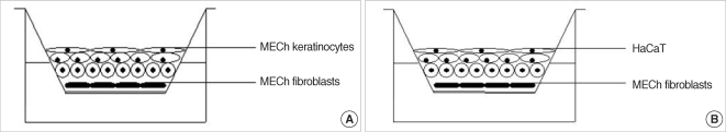
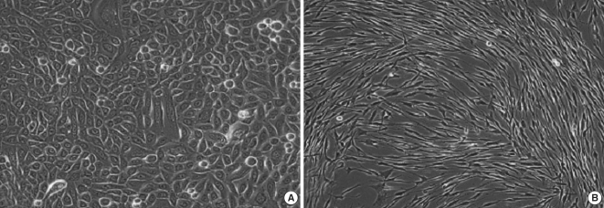
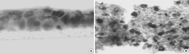
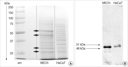
Primary keratinocytes were isolated from cholesteatoma tissue, co-cultured with preliminary prepared feeder layer from cholesteatoma fibroblasts and subsequently air-exposed. The protein profile of cholesteatoma keratinocytes and HaCaT cells was evaluated by means of immunoblot using monoclonal antibody against cytokeratin (CK) 13 and 16. Tissue localization of CK 13 and 16 was accomplished with immunohistochemistry.
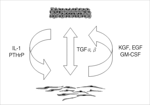
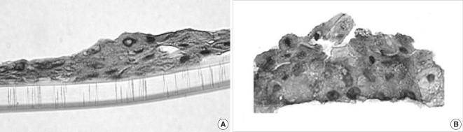
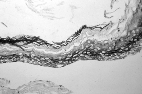
Different protein profile and stronger expression of CK 13 and 16 were demonstrated in cholesteatoma keratinocytes in comparison with HaCaT cells. Bigger stratification was observed in the 3D-in vitro systems when both cholesteatoma keratinocytes and HaCaT cells were respectively co-cultured with fibroblasts in comparison with the corresponding control groups without fibroblasts.
3D-model demonstrates the significance of intercellular interaction between components of cholesteatoma tissue.
实验模型对于研究中耳胆脂瘤的发病机制非常重要,但直到现在才建立。我们旨在使用从胆脂瘤组织中分离的原代角质形成细胞和成纤维细胞建立中耳胆脂瘤体外模型。HaCaT 细胞系被用作“皮肤等效物”,并比较胆脂瘤角质形成细胞和 HaCaT 细胞的均匀度等级。
从胆脂瘤组织中分离原代角质形成细胞,与预先制备的胆脂瘤成纤维细胞饲养层共培养,然后暴露于空气中。使用针对细胞角蛋白 (CK) 13 和 16 的单克隆抗体通过免疫印迹评估胆脂瘤角质形成细胞和 HaCaT 细胞的蛋白质谱。用免疫组织化学法完成 CK 13 和 16 的组织定位。
与 HaCaT 细胞相比,胆脂瘤角质形成细胞表现出不同的蛋白质谱和更强的 CK 13 和 16 表达。与相应的无成纤维细胞对照组相比,当胆脂瘤角质形成细胞和 HaCaT 细胞分别与成纤维细胞共培养时,3D 体外系统中观察到更大的分层。
3D 模型显示了胆脂瘤组织成分之间细胞间相互作用的重要性。