• 文献检索
  • 文档翻译
  • 深度研究
  • 学术资讯
  • Suppr Zotero 插件Zotero 插件
  • 邀请有礼
  • 套餐&价格
  • 历史记录
应用&插件
Suppr Zotero 插件Zotero 插件浏览器插件Mac 客户端Windows 客户端微信小程序
定价
高级版会员购买积分包购买API积分包
服务
文献检索文档翻译深度研究API 文档MCP 服务
关于我们
关于 Suppr公司介绍联系我们用户协议隐私条款
关注我们

Suppr 超能文献

核心技术专利:CN118964589B侵权必究
粤ICP备2023148730 号-1Suppr @ 2026

文献检索

告别复杂PubMed语法,用中文像聊天一样搜索,搜遍4000万医学文献。AI智能推荐,让科研检索更轻松。

立即免费搜索

文件翻译

保留排版,准确专业,支持PDF/Word/PPT等文件格式,支持 12+语言互译。

免费翻译文档

深度研究

AI帮你快速写综述,25分钟生成高质量综述,智能提取关键信息,辅助科研写作。

立即免费体验

左心室梗死面积、梗死周边区及乳头肌瘢痕测量:高分辨率三维与传统二维延迟钆增强心脏磁共振成像的比较

Left ventricular infarct size, peri-infarct zone, and papillary scar measurements: A comparison of high-resolution 3D and conventional 2D late gadolinium enhancement cardiac MR.

作者信息

Peters Dana C, Appelbaum Evan A, Nezafat Reza, Dokhan Basem, Han Yuchi, Kissinger Kraig V, Goddu Beth, Manning Warren J

机构信息

Beth Israel Deaconess Medical Center, Department of Medicine (Cardiovascular Division), Boston, Massachusetts, USA.

出版信息

J Magn Reson Imaging. 2009 Oct;30(4):794-800. doi: 10.1002/jmri.21897.

DOI:10.1002/jmri.21897
PMID:19787731
原文链接:https://pmc.ncbi.nlm.nih.gov/articles/PMC2981352/
Abstract

PURPOSE

To compare higher spatial resolution 3D late gadolinium enhancement (LGE) cardiovascular magnetic resonance (Cardiac MR) with 2D LGE in patients with prior myocardial infarction.

MATERIALS AND METHODS

Fourteen patients were studied using high spatial resolution 3D LGE (1.3 x 1.3 x 5.0 mm(3)) and conventional 2D LGE (2 x 2 x 8 mm(3)) scans. The signal-to-noise ratio (SNR) and contrast-to-noise ratio (CNR) were measured. Total infarct volume, peri-infarct volume measured in a limited slab, and papillary muscle scar volume were compared using Bland-Altman analysis. Image quality was graded.

RESULTS

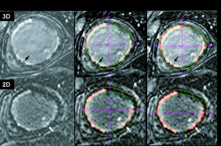
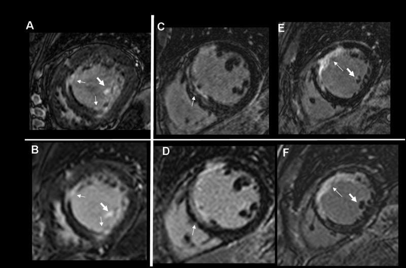
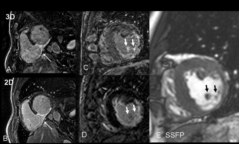
3D LGE had higher scar SNR (P < 0.001), higher myocardial SNR (P = 0.001), higher papillary scar-blood CNR (P = 0.01), and greater sharpness (P = 0.01). The scar volumes agreed (14.5 +/- 8.2 for 2D, vs. 13.2 +/- 8.8 for 3D), with bias +/- 2 standard deviations (SDs) of 0.5 +/- 6.8 mL, P = 0.59 R = 0.91. The peri-infarct volumes correlated but less strongly than scar (P = 0.40, R = 0.77). For patients with more heterogeneous scar, larger peri-infarct volumes were measured by 3D (1.9 +/- 1.1 mL for 2D vs. 2.4 +/- 1.6 mL for 3D, P = 0.15, in the matched region). Papillary scar, present in 6/14 (42%) patients, was more confidently identified on 3D LGE.

CONCLUSION

Higher spatial resolution 3D LGE provides sharper images and higher SNR, but less myocardial nulling. Scar volumes agree well, with peri-infarct volumes correlating less well. 3D LGE may be superior in visualization of papillary muscle scar.

摘要

目的

比较高空间分辨率三维延迟钆增强(LGE)心血管磁共振成像(心脏磁共振成像)与二维LGE在既往心肌梗死患者中的应用。

材料与方法

对14例患者进行了高空间分辨率三维LGE(1.3×1.3×5.0 mm³)和传统二维LGE(2×2×8 mm³)扫描。测量了信噪比(SNR)和对比噪声比(CNR)。使用Bland-Altman分析比较了总梗死体积、在有限层面测量的梗死周边体积和乳头肌瘢痕体积。对图像质量进行了分级。

结果

三维LGE具有更高的瘢痕SNR(P<0.001)、更高的心肌SNR(P = 0.001)、更高的乳头肌瘢痕-血液CNR(P = 0.01)以及更高的清晰度(P = 0.01)。瘢痕体积一致(二维为14.5±8.2,三维为13.2±8.8),偏差±2标准差(SDs)为0.5±6.8 mL,P = 0.59,R = 0.91。梗死周边体积具有相关性,但相关性不如瘢痕强(P = 0.40,R = 0.77)。对于瘢痕更不均匀的患者,三维测量的梗死周边体积更大(匹配区域中二维为1.9±1.1 mL,三维为2.4±1.6 mL,P = 0.15)。6/14(42%)的患者存在乳头肌瘢痕,在三维LGE上能更可靠地识别。

结论

高空间分辨率三维LGE提供更清晰的图像和更高的SNR,但心肌抑制效果较差。瘢痕体积一致性良好,梗死周边体积相关性较差。三维LGE在乳头肌瘢痕的可视化方面可能更具优势。

https://cdn.ncbi.nlm.nih.gov/pmc/blobs/9736/2981352/adfbfa480556/nihms185663f4.jpg
https://cdn.ncbi.nlm.nih.gov/pmc/blobs/9736/2981352/e2a772b96918/nihms185663f1.jpg
https://cdn.ncbi.nlm.nih.gov/pmc/blobs/9736/2981352/4fa5c4580e6c/nihms185663f2.jpg
https://cdn.ncbi.nlm.nih.gov/pmc/blobs/9736/2981352/13d1f9f8d734/nihms185663f3.jpg
https://cdn.ncbi.nlm.nih.gov/pmc/blobs/9736/2981352/adfbfa480556/nihms185663f4.jpg

相似文献

1
Left ventricular infarct size, peri-infarct zone, and papillary scar measurements: A comparison of high-resolution 3D and conventional 2D late gadolinium enhancement cardiac MR.左心室梗死面积、梗死周边区及乳头肌瘢痕测量:高分辨率三维与传统二维延迟钆增强心脏磁共振成像的比较
J Magn Reson Imaging. 2009 Oct;30(4):794-800. doi: 10.1002/jmri.21897.
2
Improved border sharpness of post-infarct scar by a novel self-navigated free-breathing high-resolution 3D whole-heart inversion recovery magnetic resonance approach.通过一种新型的自导航自由呼吸高分辨率三维全心反转恢复磁共振方法提高梗死灶后瘢痕的边界清晰度。
Int J Cardiovasc Imaging. 2016 Dec;32(12):1735-1744. doi: 10.1007/s10554-016-0963-4. Epub 2016 Aug 22.
3
Feasibility study of a single breath-hold, 3D mDIXON pulse sequence for late gadolinium enhancement imaging of ischemic scar.单屏气 3D mDIXON 脉冲序列用于缺血性瘢痕晚期钆增强成像的可行性研究。
J Magn Reson Imaging. 2019 May;49(5):1437-1445. doi: 10.1002/jmri.26519. Epub 2018 Dec 31.
4
Assessment of left ventricular myocardial scar in coronary artery disease by a three-dimensional MR imaging technique.应用三维磁共振成像技术评估冠状动脉疾病中的左心室心肌瘢痕。
J Magn Reson Imaging. 2013 Jul;38(1):72-9. doi: 10.1002/jmri.23962. Epub 2012 Dec 7.
5
High spatial resolution free-breathing 3D late gadolinium enhancement cardiac magnetic resonance imaging in ischaemic and non-ischaemic cardiomyopathy: quantitative assessment of scar mass and image quality.高空间分辨率自由呼吸 3D 晚期钆增强心脏磁共振成像在缺血性和非缺血性心肌病中的应用:瘢痕质量和图像质量的定量评估。
Eur Radiol. 2018 Sep;28(9):4027-4035. doi: 10.1007/s00330-018-5361-y. Epub 2018 Apr 6.
6
Acute, subacute, and chronic myocardial infarction: quantitative comparison of 2D and 3D late gadolinium enhancement MR imaging.急性、亚急性和慢性心肌梗死:二维和三维晚期钆增强磁共振成像的定量比较。
Radiology. 2011 Jun;259(3):704-11. doi: 10.1148/radiol.11102216. Epub 2011 Apr 5.
7
Gray blood late gadolinium enhancement cardiovascular magnetic resonance for improved detection of myocardial scar.钆延迟增强心血管磁共振检测心肌瘢痕的灰色血液。
J Cardiovasc Magn Reson. 2018 Mar 22;20(1):22. doi: 10.1186/s12968-018-0442-2.
8
Myocardial Scar Detection Using High-Resolution Free-Breathing 3D Dark-Blood and Standard Breath-Holding 2D Bright-Blood Late Gadolinium Enhancement MRI: A Comparison of Observer Confidence.高分辨率自由呼吸 3D 黑血和标准屏气 2D 亮血钆延迟增强 MRI 检测心肌瘢痕:观察者信心的比较。
Top Magn Reson Imaging. 2023 Jun 1;32(3):27-32. doi: 10.1097/RMR.0000000000000304. Epub 2023 Apr 16.
9
Infarct quantification using 3D inversion recovery and 2D phase sensitive inversion recovery; validation in patients and ex vivo.使用 3D 反转恢复和 2D 相位敏感反转恢复进行梗塞定量;在患者和离体中的验证。
BMC Cardiovasc Disord. 2013 Dec 5;13:110. doi: 10.1186/1471-2261-13-110.
10
3D late gadolinium enhanced cardiovascular MR with CENTRA-PLUS profile/view ordering: Feasibility of right ventricular myocardial damage assessment using a swine animal model.采用CENTRA-PLUS轮廓/视图排序的3D延迟钆增强心血管磁共振成像:使用猪动物模型评估右心室心肌损伤的可行性。
Magn Reson Imaging. 2017 Jun;39:7-14. doi: 10.1016/j.mri.2017.01.015. Epub 2017 Jan 25.

引用本文的文献

1
Late Gadolinium Enhancement of Nonischemic Cardiomyopathy at 5.0 T versus 3.0 T: A Crossover Design Study.5.0T与3.0T场强下非缺血性心肌病的延迟钆增强:一项交叉设计研究
Radiol Cardiothorac Imaging. 2024 Dec;6(6):e240035. doi: 10.1148/ryct.240035.
2
Compressed SENSE accelerated 3D single-breath-hold late gadolinium enhancement cardiovascular magnetic resonance with isotropic resolution: clinical evaluation.具有各向同性分辨率的压缩感知加速三维屏气晚期钆增强心血管磁共振成像:临床评估
Front Cardiovasc Med. 2023 Nov 30;10:1305649. doi: 10.3389/fcvm.2023.1305649. eCollection 2023.
3
Diagnostic and prognostic role of late gadolinium enhancement in cardiomyopathies.

本文引用的文献

1
Cardiovascular magnetic resonance characterization of mitral valve prolapse.二尖瓣脱垂的心血管磁共振特征
JACC Cardiovasc Imaging. 2008 May;1(3):294-303. doi: 10.1016/j.jcmg.2008.01.013.
2
Ventricular arrhythmias originating from a papillary muscle in patients without prior infarction: a comparison with fascicular arrhythmias.无既往梗死患者源自乳头肌的室性心律失常:与束支心律失常的比较
Heart Rhythm. 2008 Nov;5(11):1530-7. doi: 10.1016/j.hrthm.2008.08.032. Epub 2008 Sep 3.
3
Post-infarction ventricular arrhythmias originating in papillary muscles.
延迟钆增强在心肌病中的诊断和预后作用。
Eur Heart J Suppl. 2023 Apr 26;25(Suppl C):C130-C136. doi: 10.1093/eurheartjsupp/suad015. eCollection 2023 May.
4
Image Quality of Submillimeter High-Spatial-Resolution 2D Late Gadolinium-enhanced Images in Cardiac MRI: A Feasibility Study.心脏磁共振成像中高空间分辨率亚毫米二维延迟钆增强图像的图像质量:一项可行性研究。
Radiol Cardiothorac Imaging. 2022 Dec 8;4(6):e220111. doi: 10.1148/ryct.220111. eCollection 2022 Dec.
5
Myocardial scar detection in free-breathing Dixon-based fat- and water-separated 3D inversion recovery late-gadolinium enhancement whole heart MRI.自由呼吸 Dixon 基的脂肪和水分离 3D 反转恢复晚期钆增强全心 MRI 中的心肌瘢痕检测。
Int J Cardiovasc Imaging. 2023 Jan;39(1):135-144. doi: 10.1007/s10554-022-02701-0. Epub 2022 Aug 23.
6
Assessment of Papillary Muscle Infarction with Dark-Blood Delayed Enhancement Cardiac MRI in Canines and Humans.犬和人类心脏黑血延迟增强 MRI 评估乳头肌梗死。
Radiology. 2022 Nov;305(2):329-338. doi: 10.1148/radiol.220251. Epub 2022 Jul 26.
7
Ablation Lesion Assessment with MRI.利用磁共振成像评估消融病灶
Arrhythm Electrophysiol Rev. 2022 Apr;11:e02. doi: 10.15420/aer.2021.63.
8
Whole-Heart High-Resolution Late Gadolinium Enhancement: Techniques and Clinical Applications.全心高分辨率晚期钆增强:技术和临床应用。
J Magn Reson Imaging. 2022 Apr;55(4):967-987. doi: 10.1002/jmri.27732. Epub 2021 Jun 21.
9
Comparison of 3D and 2D late gadolinium enhancement magnetic resonance imaging in patients with acute and chronic myocarditis.比较 3D 和 2D 晚期钆增强磁共振成像在急性和慢性心肌炎患者中的应用。
Int J Cardiovasc Imaging. 2021 Jan;37(1):305-313. doi: 10.1007/s10554-020-01966-7. Epub 2020 Aug 13.
10
Motion-corrected 3D whole-heart water-fat high-resolution late gadolinium enhancement cardiovascular magnetic resonance imaging.运动校正的 3D 全心水脂高分辨率晚期钆增强心血管磁共振成像。
J Cardiovasc Magn Reson. 2020 Jul 20;22(1):53. doi: 10.1186/s12968-020-00649-5.
起源于乳头肌的心肌梗死后室性心律失常。
J Am Coll Cardiol. 2008 May 6;51(18):1794-802. doi: 10.1016/j.jacc.2008.01.046.
4
Significance of papillary muscle abnormalities identified by cardiovascular magnetic resonance in hypertrophic cardiomyopathy.心血管磁共振识别的肥厚型心肌病乳头肌异常的意义
Am J Cardiol. 2008 Mar 1;101(5):668-73. doi: 10.1016/j.amjcard.2007.10.032. Epub 2007 Dec 21.
5
A fast navigator-gated 3D sequence for delayed enhancement MRI of the myocardium: comparison with breathhold 2D imaging.一种用于心肌延迟强化磁共振成像的快速导航门控3D序列:与屏气2D成像的比较。
J Magn Reson Imaging. 2008 Apr;27(4):802-8. doi: 10.1002/jmri.21296.
6
Infarct tissue heterogeneity by magnetic resonance imaging identifies enhanced cardiac arrhythmia susceptibility in patients with left ventricular dysfunction.通过磁共振成像检测到的梗死组织异质性可识别左心室功能障碍患者增强的心律失常易感性。
Circulation. 2007 Apr 17;115(15):2006-14. doi: 10.1161/CIRCULATIONAHA.106.653568. Epub 2007 Mar 26.
7
Characterization of the peri-infarct zone by contrast-enhanced cardiac magnetic resonance imaging is a powerful predictor of post-myocardial infarction mortality.通过对比增强心脏磁共振成像对梗死周边区进行特征描述是心肌梗死后死亡率的有力预测指标。
Circulation. 2006 Jul 4;114(1):32-9. doi: 10.1161/CIRCULATIONAHA.106.613414. Epub 2006 Jun 26.
8
Impact of unrecognized myocardial scar detected by cardiac magnetic resonance imaging on event-free survival in patients presenting with signs or symptoms of coronary artery disease.心脏磁共振成像检测到的未被识别的心肌瘢痕对出现冠状动脉疾病体征或症状患者无事件生存期的影响。
Circulation. 2006 Jun 13;113(23):2733-43. doi: 10.1161/CIRCULATIONAHA.105.570648. Epub 2006 Jun 5.
9
Myocardial viability: assessment with three-dimensional MR imaging in pigs and patients.心肌存活:猪和患者的三维磁共振成像评估
Radiology. 2006 Jun;239(3):703-9. doi: 10.1148/radiol.2393050586. Epub 2006 Apr 26.
10
Magnetic resonance assessment of the substrate for inducible ventricular tachycardia in nonischemic cardiomyopathy.非缺血性心肌病中可诱导室性心动过速底物的磁共振评估
Circulation. 2005 Nov 1;112(18):2821-5. doi: 10.1161/CIRCULATIONAHA.105.549659.