Messiou C, Cook G, deSouza N M
Cancer Research UK Clinical Magnetic Resonance Research Group, Institute of Cancer Research and Royal Marsden NHS Foundation Trust, Surrey, UK.
Br J Cancer. 2009 Oct 20;101(8):1225-32. doi: 10.1038/sj.bjc.6605334. Epub 2009 Sep 29.
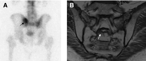
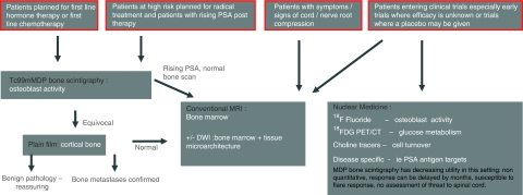
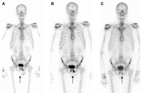
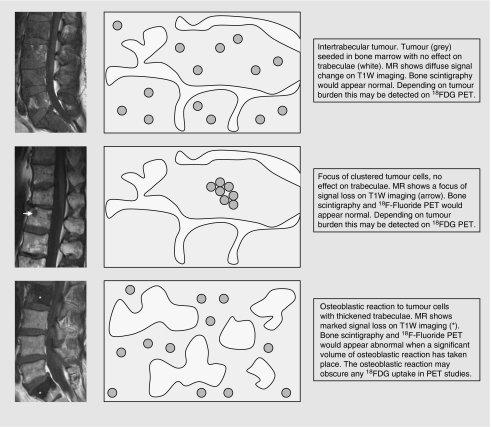
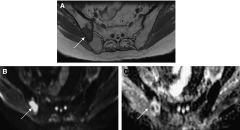
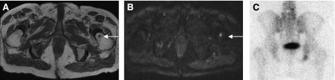
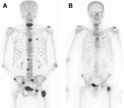
Imaging bone metastases from prostate cancer presents several challenges. The lesions are usually sclerotic and appear late on the conventional X-ray. Bone scintigraphy is the mainstay of lesion detection, but is often not suitable for assessment of treatment response, particularly because of a 'flare' phenomenon after therapy. Magnetic resonance imaging is increasingly used in assessment, and newer techniques allow quantitation. In addition to (18)F-fluorodeoxyglucose ((18)FDG), newer PET isotopes are also showing promise in lesion detection and response assessment. This article reviews the available imaging modalities for evaluating prostatic bony metastases, and links them to the underlying pathological changes within bone lesions.
前列腺癌骨转移的影像学检查存在诸多挑战。这些病变通常为硬化性,在传统X射线上出现较晚。骨闪烁扫描是检测病变的主要方法,但通常不适用于评估治疗反应,尤其是由于治疗后会出现“闪烁”现象。磁共振成像在评估中越来越常用,新技术能够进行定量分析。除了(18)F-氟脱氧葡萄糖((18)FDG),新型PET同位素在病变检测和反应评估中也显示出前景。本文综述了用于评估前列腺骨转移的现有影像学方法,并将它们与骨病变内的潜在病理变化联系起来。