Department of Radiology and Imaging Sciences, National Institutes of Health, Clinical Center, Bethesda, MD, USA.
AJR Am J Roentgenol. 2009 Dec;193(6):1500-3. doi: 10.2214/AJR.09.3365.
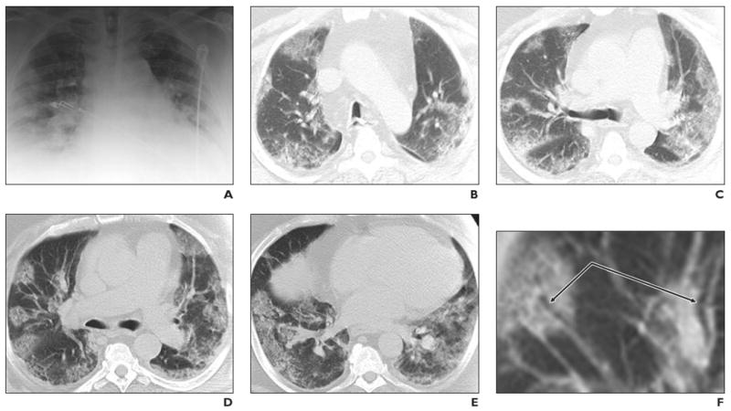
Although most cases of swine-origin influenza A (H1N1) virus (S-OIV) have been self-limited, fatal cases raise questions about virulence and radiology's role in early detection. We describe the radiographic and CT findings in a fatal S-OIV infection.
Radiography showed peripheral lung opacities. CT revealed peripheral ground-glass opacities suggesting peribronchial injury. These imaging findings raised suspicion of S-OIV despite negative H1N1 influenza rapid antigen test results from two nasopharyngeal swabs; subsequently, those results were proven to be false-negatives by reverse transcriptase polymerase chain reaction. This case suggests a role for CT in the early recognition of severe S-OIV.
尽管大多数猪源甲型 H1N1 流感病毒(S-OIV)感染病例为自限性,但致死病例引发了关于其毒力和放射学在早期检测中作用的疑问。我们描述了一例致死性 S-OIV 感染的放射学和 CT 表现。
X 线表现为外周肺实变。CT 显示外周磨玻璃影,提示支气管周围损伤。尽管两次鼻咽拭子的 H1N1 流感快速抗原检测结果均为阴性,但这些影像学表现仍提示存在 S-OIV 感染的可能;随后,逆转录聚合酶链反应证实这些结果为假阴性。本病例提示 CT 在早期识别重症 S-OIV 中具有一定作用。