Department of Radiology, University of Pennsylvania, Philadelphia, PA 19104, USA.
Neuroimage. 2010 Apr 1;50(2):434-45. doi: 10.1016/j.neuroimage.2009.12.007. Epub 2009 Dec 31.
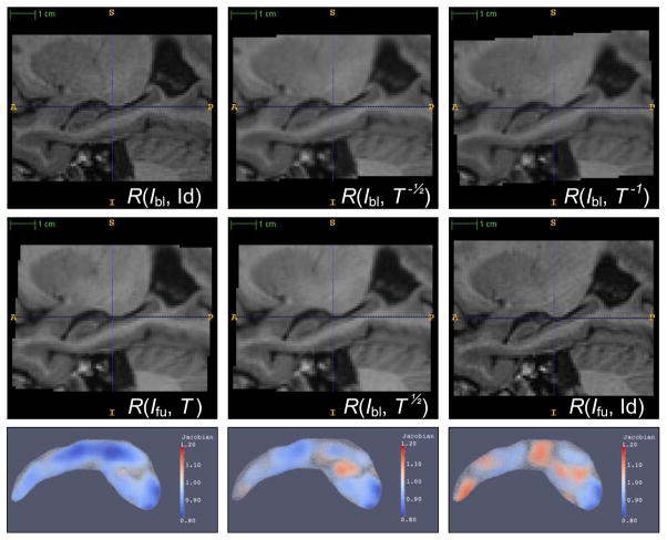
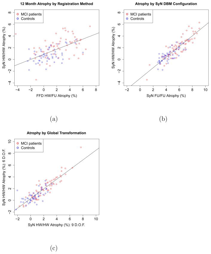
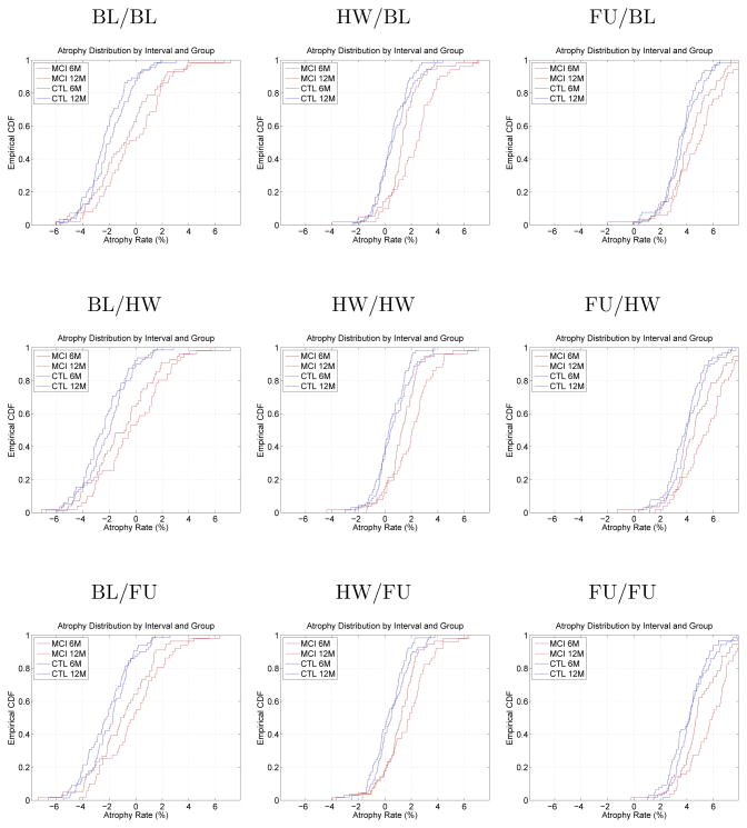
Measurement of brain change due to neurodegenerative disease and treatment is one of the fundamental tasks of neuroimaging. Deformation-based morphometry (DBM) has been long recognized as an effective and sensitive tool for estimating the change in the volume of brain regions over time. This paper demonstrates that a straightforward application of DBM to estimate the change in the volume of the hippocampus can result in substantial bias, i.e., an overestimation of the rate of change in hippocampal volume. In ADNI data, this bias is manifested as a non-zero intercept of the regression line fitted to the 6 and 12 month rates of hippocampal atrophy. The bias is further confirmed by applying DBM to repeat scans of subjects acquired on the same day. This bias appears to be the result of asymmetry in the interpolation of baseline and followup images during longitudinal image registration. Correcting this asymmetry leads to bias-free atrophy estimation.
测量因神经退行性疾病和治疗而导致的大脑变化是神经影像学的基本任务之一。基于变形的形态计量学(DBM)一直被认为是估计脑区随时间变化的体积的有效且敏感的工具。本文表明,直接应用 DBM 来估计海马体体积的变化会导致显著的偏差,即高估了海马体体积变化的速度。在 ADNI 数据中,这种偏差表现为拟合至 6 个月和 12 个月海马体萎缩率的回归线上的非零截距。通过将 DBM 应用于同一天获取的受试者的重复扫描进一步证实了这种偏差。这种偏差似乎是在纵向图像配准过程中对基线和随访图像进行插值时的不对称性造成的。纠正这种不对称性可导致无偏差的萎缩估计。