• 文献检索
  • 文档翻译
  • 深度研究
  • 学术资讯
  • Suppr Zotero 插件Zotero 插件
  • 邀请有礼
  • 套餐&价格
  • 历史记录
应用&插件
Suppr Zotero 插件Zotero 插件浏览器插件Mac 客户端Windows 客户端微信小程序
定价
高级版会员购买积分包购买API积分包
服务
文献检索文档翻译深度研究API 文档MCP 服务
关于我们
关于 Suppr公司介绍联系我们用户协议隐私条款
关注我们

Suppr 超能文献

核心技术专利:CN118964589B侵权必究
粤ICP备2023148730 号-1Suppr @ 2026

文献检索

告别复杂PubMed语法,用中文像聊天一样搜索,搜遍4000万医学文献。AI智能推荐,让科研检索更轻松。

立即免费搜索

文件翻译

保留排版,准确专业,支持PDF/Word/PPT等文件格式,支持 12+语言互译。

免费翻译文档

深度研究

AI帮你快速写综述,25分钟生成高质量综述,智能提取关键信息,辅助科研写作。

立即免费体验

共配准光学相干断层扫描和荧光分子成像用于同时进行形态学和分子成像。

Co-registered optical coherence tomography and fluorescence molecular imaging for simultaneous morphological and molecular imaging.

机构信息

Fischell Department of Bioengineering, University of Maryland, College Park, MD 20742, USA.

出版信息

Phys Med Biol. 2010 Jan 7;55(1):191-206. doi: 10.1088/0031-9155/55/1/011.

DOI:10.1088/0031-9155/55/1/011
PMID:20009192
原文链接:https://pmc.ncbi.nlm.nih.gov/articles/PMC2951762/
Abstract

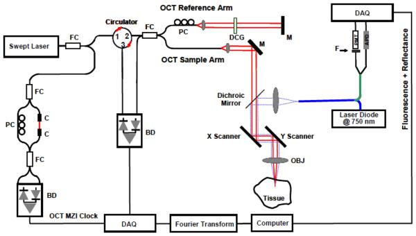
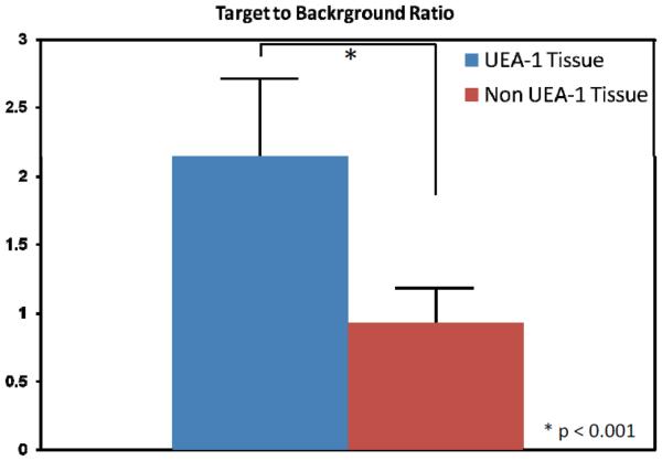
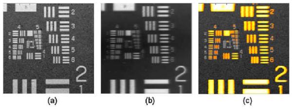
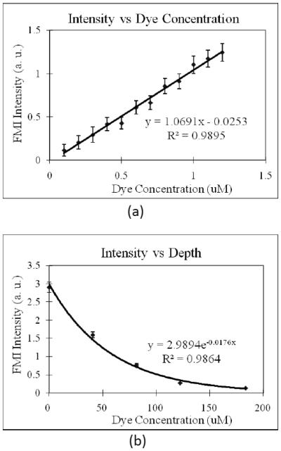
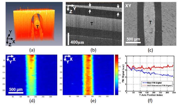
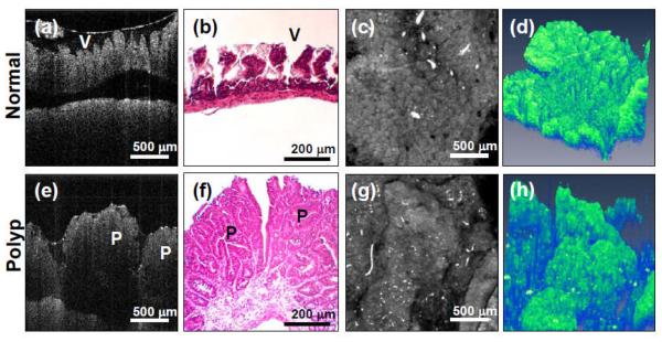
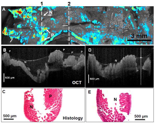
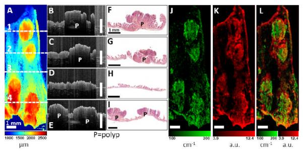
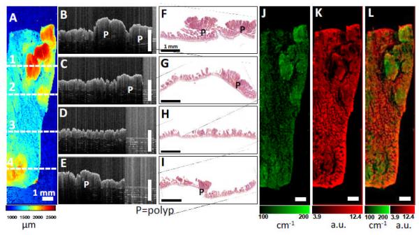
Optical coherence tomography (OCT) provides high-resolution, cross-sectional imaging of tissue microstructure in situ and in real time, while fluorescence molecular imaging (FMI) enables the visualization of basic molecular processes. There is a great deal of interest in combining these two modalities so that the tissue's structural and molecular information can be obtained simultaneously. This could greatly benefit biomedical applications such as detecting early diseases and monitoring therapeutic interventions. In this research, an optical system that combines OCT and FMI was developed. The system demonstrated that it could co-register en face OCT and FMI images with a 2.4 x 2.4 mm(2) field-of-view. The transverse resolutions of OCT and FMI of the system are both approximately 10 microm. Capillary tubes filled with fluorescent dye Cy 5.5 in different concentrations under a scattering medium are used as the phantom. En face OCT images of the phantoms were obtained and successfully co-registered with FMI images that were acquired simultaneously. A linear relationship between FMI intensity and dye concentration was observed. The relationship between FMI intensity and target fluorescence tube depth measured by OCT images was also observed and compared with theoretical modeling. This relationship could help in correcting reconstructed dye concentration. Imaging of colon polyps of the APC(min) mouse model is presented as an example of biological applications of this co-registered OCT/FMI system.

摘要

光学相干断层扫描(OCT)提供组织微观结构的高分辨率、横截面成像,实时原位,而荧光分子成像(FMI)能够可视化基本的分子过程。人们对将这两种模式结合起来非常感兴趣,以便同时获得组织的结构和分子信息。这将极大地有益于生物医学应用,如早期疾病检测和治疗干预监测。在这项研究中,开发了一种结合 OCT 和 FMI 的光学系统。该系统证明它可以以 2.4 x 2.4 毫米(2)的视场共注册 OCT 和 FMI 图像。该系统的 OCT 和 FMI 的横向分辨率均约为 10 微米。在散射介质下,使用充满不同浓度荧光染料 Cy 5.5 的毛细管作为模型。获得了模型的 OCT 图像,并与同时获得的 FMI 图像成功地共注册。观察到 FMI 强度与染料浓度之间存在线性关系。还观察到并比较了 OCT 图像测量的 FMI 强度与目标荧光管深度之间的关系,并与理论建模进行了比较。这种关系有助于校正重建的染料浓度。以 APC(min) 小鼠模型的结肠息肉成像为例,展示了这种共注册的 OCT/FMI 系统的生物应用。

https://cdn.ncbi.nlm.nih.gov/pmc/blobs/4e98/2951762/24a1114745de/nihms-233002-f0009.jpg
https://cdn.ncbi.nlm.nih.gov/pmc/blobs/4e98/2951762/bf1bdc0bbc0e/nihms-233002-f0001.jpg
https://cdn.ncbi.nlm.nih.gov/pmc/blobs/4e98/2951762/db6d0853b9d5/nihms-233002-f0002.jpg
https://cdn.ncbi.nlm.nih.gov/pmc/blobs/4e98/2951762/c0dbf952282c/nihms-233002-f0003.jpg
https://cdn.ncbi.nlm.nih.gov/pmc/blobs/4e98/2951762/de2b5a0fab17/nihms-233002-f0004.jpg
https://cdn.ncbi.nlm.nih.gov/pmc/blobs/4e98/2951762/6f2625ab850b/nihms-233002-f0005.jpg
https://cdn.ncbi.nlm.nih.gov/pmc/blobs/4e98/2951762/21b70a80b570/nihms-233002-f0006.jpg
https://cdn.ncbi.nlm.nih.gov/pmc/blobs/4e98/2951762/6eb13f647acc/nihms-233002-f0007.jpg
https://cdn.ncbi.nlm.nih.gov/pmc/blobs/4e98/2951762/1826c9788182/nihms-233002-f0008.jpg
https://cdn.ncbi.nlm.nih.gov/pmc/blobs/4e98/2951762/24a1114745de/nihms-233002-f0009.jpg

相似文献

1
Co-registered optical coherence tomography and fluorescence molecular imaging for simultaneous morphological and molecular imaging.共配准光学相干断层扫描和荧光分子成像用于同时进行形态学和分子成像。
Phys Med Biol. 2010 Jan 7;55(1):191-206. doi: 10.1088/0031-9155/55/1/011.
2
Three-dimensional coregistered optical coherence tomography and line-scanning fluorescence laminar optical tomography.三维配准光学相干断层扫描与线扫描荧光层状光学断层扫描。
Opt Lett. 2009 Jun 1;34(11):1615-7. doi: 10.1364/ol.34.001615.
3
Three-dimensional retinal imaging with high-speed ultrahigh-resolution optical coherence tomography.采用高速超高分辨率光学相干断层扫描技术的三维视网膜成像
Ophthalmology. 2005 Oct;112(10):1734-46. doi: 10.1016/j.ophtha.2005.05.023.
4
Multiscale multimodal imaging with multiphoton microscopy and optical coherence tomography.多光子显微镜和光相干断层扫描的多尺度多模态成像。
Opt Lett. 2011 Dec 15;36(24):4800-2. doi: 10.1364/OL.36.004800.
5
Microscope integrated optical coherence tomography system combined with augmented reality.显微镜集成光学相干断层扫描系统结合增强现实。
Opt Express. 2021 Mar 15;29(6):9407-9418. doi: 10.1364/OE.420375.
6
Investigations of the eye fundus using a simultaneous optical coherence tomography/indocyanine green fluorescence imaging system.使用同步光学相干断层扫描/吲哚菁绿荧光成像系统对眼底进行研究。
J Biomed Opt. 2007 Jan-Feb;12(1):014019. doi: 10.1117/1.2434970.
7
High-resolution three-dimensional imaging of biofilm development using optical coherence tomography.使用光学相干断层扫描技术对生物膜形成进行高分辨率三维成像。
J Biomed Opt. 2006 May-Jun;11(3):34001. doi: 10.1117/1.2209962.
8
Tissue perfusion modelling in optical coherence tomography.光学相干断层扫描中的组织灌注建模
Biomed Eng Online. 2017 Feb 8;16(1):27. doi: 10.1186/s12938-017-0320-4.
9
In vivo, dual-modality OCT/LIF imaging using a novel VEGF receptor-targeted NIR fluorescent probe in the AOM-treated mouse model.体内,使用新型 VEGF 受体靶向近红外荧光探针在 AOM 处理的小鼠模型中进行的双模态 OCT/LIF 成像。
Mol Imaging Biol. 2011 Dec;13(6):1173-82. doi: 10.1007/s11307-010-0450-6.
10
Combined multiphoton microscopy and optical coherence tomography using a 12-fs broadband source.使用12飞秒宽带光源的联合多光子显微镜和光学相干断层扫描技术。
J Biomed Opt. 2006 Mar-Apr;11(2):020502. doi: 10.1117/1.2193428.

引用本文的文献

1
Retinal ganglion cell repopulation for vision restoration in optic neuropathy: a roadmap from the RReSTORe Consortium.视网膜神经节细胞再灌注以恢复视神经病变的视力:RReSTORe 联盟的路线图。
Mol Neurodegener. 2023 Sep 21;18(1):64. doi: 10.1186/s13024-023-00655-y.
2
Dual-modality imaging system for monitoring human heart organoids beating in vitro.用于监测体外人类心脏类器官跳动的双模式成像系统。
Opt Lett. 2023 Aug 1;48(15):3929-3932. doi: 10.1364/OL.493824.
3
Nanotheranostic Strategies for Cancer Immunotherapy.用于癌症免疫治疗的纳米诊疗策略。

本文引用的文献

1
Refractive indices of water and ice in the 0.65- to 2.5-µm spectral range.0.65至2.5微米光谱范围内水和冰的折射率。
Appl Opt. 1993 Jul 1;32(19):3531-40. doi: 10.1364/AO.32.003531.
2
Light scattering in Intralipid-10% in the wavelength range of 400-1100 nm.10% 英脱利匹特在400 - 1100纳米波长范围内的光散射
Appl Opt. 1991 Nov 1;30(31):4507-14. doi: 10.1364/AO.30.004507.
3
Optical properties of rat liver between 350 and 2200 nm.350至2200纳米波长范围内大鼠肝脏的光学特性。
Small Methods. 2022 Dec;6(12):e2200718. doi: 10.1002/smtd.202200718. Epub 2022 Nov 16.
4
Mapping Lymph Node during Indocyanine Green Fluorescence-Imaging Guided Gastric Oncologic Surgery: Current Applications and Future Directions.吲哚菁绿荧光成像引导下胃癌手术中的淋巴结定位:当前应用与未来方向
Cancers (Basel). 2022 Oct 20;14(20):5143. doi: 10.3390/cancers14205143.
5
Design and synthesis of lipid-mimetic cationic iridium complexes and their liposomal formulation for and application in luminescent bioimaging.脂质模拟阳离子铱配合物的设计与合成及其脂质体制剂在发光生物成像中的应用
RSC Adv. 2020 Apr 8;10(24):14431-14440. doi: 10.1039/d0ra01114b. eCollection 2020 Apr 6.
6
Mesoporous Silica-Coated Gold Nanoparticles for Multimodal Imaging and Reactive Oxygen Species Sensing of Stem Cells.用于干细胞多模态成像和活性氧传感的介孔二氧化硅包覆金纳米颗粒
ACS Appl Nano Mater. 2022 Mar 25;5(3):3237-3251. doi: 10.1021/acsanm.1c03640. Epub 2022 Mar 14.
7
Advances in Endoscopic Photoacoustic Imaging.内镜光声成像的进展
Photonics. 2021 Jul;8(7). doi: 10.3390/photonics8070281. Epub 2021 Jul 16.
8
Rapid pseudo-H&E imaging using a fluorescence-inbuilt optical coherence microscopic imaging system.使用内置荧光的光学相干显微成像系统进行快速伪苏木精-伊红成像。
Biomed Opt Express. 2021 Jul 21;12(8):5139-5158. doi: 10.1364/BOE.431586. eCollection 2021 Aug 1.
9
Fiber-based photoacoustic remote sensing microscopy and spectral-domain optical coherence tomography with a dual-function 1050-nm interrogation source.基于光纤的光声远程传感显微镜和光谱域光学相干断层扫描,采用双功能 1050nm 询问光源。
J Biomed Opt. 2021 Jun;26(6). doi: 10.1117/1.JBO.26.6.066502.
10
Colon phantoms with cancer lesions for endoscopic characterization with optical coherence tomography.用于光学相干断层扫描内镜特征分析的带有癌症病变的结肠模型。
Biomed Opt Express. 2021 Jan 20;12(2):955-968. doi: 10.1364/BOE.402081. eCollection 2021 Feb 1.
Appl Opt. 1989 Jun 15;28(12):2325-30. doi: 10.1364/AO.28.002325.
4
Refractive index of some mammalian tissues using a fiber optic cladding method.使用光纤包层法测量某些哺乳动物组织的折射率
Appl Opt. 1989 Jun 15;28(12):2297-303. doi: 10.1364/AO.28.002297.
5
Development of fluorescent materials for Diffuse Fluorescence Tomography standards and phantoms.用于漫反射荧光层析成像标准和体模的荧光材料的开发。
Opt Express. 2007 Jul 9;15(14):8681-94. doi: 10.1364/oe.15.008681.
6
In vivo endoscopic OCT imaging of precancer and cancer states of human mucosa.人体黏膜癌前和癌变状态的体内内镜光学相干断层扫描成像
Opt Express. 1997 Dec 22;1(13):432-40. doi: 10.1364/oe.1.000432.
7
Three-dimensional endomicroscopy of the human colon using optical coherence tomography.使用光学相干断层扫描对人体结肠进行三维内镜检查。
Opt Express. 2009 Jan 19;17(2):784-96. doi: 10.1364/oe.17.000784.
8
Automated quantification of colonic crypt morphology using integrated microscopy and optical coherence tomography.
J Biomed Opt. 2008 Sep-Oct;13(5):054055. doi: 10.1117/1.2993323.
9
Assessment of early demineralization in teeth using the signal attenuation in optical coherence tomography images.利用光学相干断层扫描图像中的信号衰减评估牙齿早期脱矿情况。
J Biomed Opt. 2008 Sep-Oct;13(5):054053. doi: 10.1117/1.2992129.
10
Simultaneous OCT/SLO/ICG imaging.光学相干断层扫描/扫描激光检眼镜/吲哚菁绿同步成像
Invest Ophthalmol Vis Sci. 2009 Feb;50(2):851-60. doi: 10.1167/iovs.08-1855. Epub 2008 Oct 24.